A team of over 150 scientists has achieved what once seemed impossible: a complete wiring and activity map of a tiny section of a mammalian brain.
This feat, part of the MICrONS Project, rivals the Human Genome Project in ambition and scope, using cutting-edge AI, microscopy, and teamwork to map over 200,000 brain cells and millions of synapses. Among many revelations, researchers uncovered surprising new rules for how inhibitory neurons selectively influence others, providing insight into how thought, memory, and disorders like Alzheimer’s might emerge from cellular interactions. This achievement opens the door to a new era in brain science and medical breakthroughs.
Cracking the Brain’s Wiring Code
From a tiny piece of brain tissue no larger than a grain of sand, scientists have achieved what once seemed impossible: creating a detailed, functional wiring diagram of part of the brain. In 1979, molecular biologist Francis Crick famously predicted it would be “[impossible] to create an exact wiring diagram for a cubic millimeter of brain tissue and the way all its neurons are firing.” But now, after seven years of work, more than 150 scientists and researchers from around the world have brought that vision much closer to reality.

MICrONS Project: A Historic Brain Mapping Breakthrough
The Machine Intelligence from Cortical Networks (MICrONS) Project has produced the most detailed wiring diagram of a mammalian brain ever made. The results, shared on April 9 across ten scientific papers published in the Nature family of journals, offer unprecedented insight into how the brain is structured and how it functions, especially within the visual system. The dataset, available through the MICrONS Explorer, totals 1.6 petabytes – about the same as 22 years of continuous HD video.
“The MICrONS advances published in this special issue of Nature are a watershed moment for neuroscience, comparable to the Human Genome Project in their transformative potential,” said David A. Markowitz, Ph.D., former IARPA program manager who coordinated this work.
“IARPA’s moonshot investment in the MICrONS program has shattered previous technological limitations, creating the first platform to study the relationship between neural structure and function at scales necessary to understand intelligence. This achievement validates our focused research approach and sets the stage for future scaling to the whole brain level.”
Building the Brain’s 3D Blueprint
Scientists at Baylor College of Medicine began by using specialized microscopes to record the brain activity from a one cubic millimeter portion of a mouse’s visual cortex as the animal watched various movies and YouTube clips. Afterward, Allen Institute researchers took that same cubic millimeter of the brain and sliced it into more than 25,000 layers, each 1/400th the width of a human hair, and used an array of electron microscopes to take high-resolution pictures of each slice. Finally, another team at Princeton University used artificial intelligence and machine learning to reconstruct the cells and connections into a 3D volume.
Combined with the recordings of brain activity, the result is the largest wiring diagram and functional map of the brain to date, containing more than 200,000 cells, four kilometers of axons (the branches that reach out to other cells), and 523 million synapses (the connection points between cells).
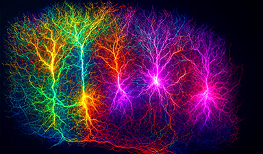
A Forest of Neural Connections
“Inside that tiny speck is an entire architecture like an exquisite forest,” said Clay Reid, M.D., Ph.D., senior investigator and one of the early founders of electron microscopy connectomics who brought this area of science to the Allen Institute 13 years ago. “It has all sorts of rules of connections that we knew from various parts of neuroscience, and within the reconstruction itself, we can test the old theories and hope to find new things that no one has ever seen before.”
The findings from the studies reveal new cell types, characteristics, organizational and functional principles, and a new way to classify cells. Among the most surprising findings was the discovery of a new principle of inhibition within the brain. Scientists previously thought of inhibitory cells – those that suppress neural activity – as a simple force that dampens the action of other cells.
However, researchers discovered a far more sophisticated level of communication: Inhibitory cells are not random in their actions; instead, they are highly selective about which excitatory cells they target, creating a network-wide system of coordination and cooperation. Some inhibitory cells work together, suppressing multiple excitatory cells, while others are more precise, targeting only specific types.
Surprising Discoveries in Brain Inhibition
“This is the future in many ways,” explained Andreas Tolias, Ph.D., one of the lead scientists who worked on this project at both Baylor College of Medicine and Stanford University. “MICrONS will stand as a landmark where we build brain foundation models that span many levels of analysis, beginning from the behavioral level to the representational level of neural activity and even to the molecular level.”
What this Means for Science and Medicine
Understanding the brain’s form and function and the ability to analyze the detailed connections between neurons at an unprecedented scale opens new possibilities for studying the brain and intelligence. It also has implications for disorders like Alzheimer’s, Parkinson’s, autism, and schizophrenia involving disruptions in neural communication.
“If you have a broken radio and you have the circuit diagram, you’ll be in a better position to fix it,” said Nuno da Costa, Ph.D., associate investigator at the Allen Institute. “We are describing a kind of Google map or blueprint of this grain of sand. In the future, we can use this to compare the brain wiring in a healthy mouse to the brain wiring in a model of disease.”
Big Science, Big Collaboration
The MICrONS Project is a collaborative effort of more than 150 scientists and researchers from the Allen Institute, Princeton, Harvard, Baylor College of Medicine, Stanford, and many others.
“Doing this kind of large, team-scale science requires a lot of cooperation,” said Forrest Collman, Ph.D., associate director of data and technology at the Allen Institute. “It requires people to dream big and to agree to tackle problems that aren’t obviously solvable, and that’s how advances happen.”
The collaborative, global effort was made possible by support from the Intelligence Advanced Research Projects Activity (IARPA) and National Institutes of Health’s Brain Research Through Advancing Innovative Neurotechnologies® Initiative, or The BRAIN Initiative®.
Foundations for Future Treatments
“The BRAIN Initiative plays a critical role in bringing together scientists from various disciplines to perform complex and challenging research that cannot be achieved in isolation,” said John Ngai, Ph.D., director of The BRAIN Initiative®. “Basic science building blocks, like how the brain is wired, are the foundation we need to better understand brain injury and disease, to bring treatments and cures closer to clinical use.”
A map of neuronal connectivity, form, and function from a grain of sand-sized portion of the brain is not just a scientific marvel, but a step toward understanding the elusive origins of thought, emotion, and consciousness. The “impossible” task first envisioned by Francis Crick in 1979 is now one step closer to reality.
Reference: “Functional connectomics spanning multiple areas of mouse visual cortex” by The MICrONS Consortium, 9 April 2025, Nature.
DOI: 10.1038/s41586-025-08790-w
News
A Grain of Brain, 523 Million Synapses, Most Complicated Neuroscience Experiment Ever Attempted
A team of over 150 scientists has achieved what once seemed impossible: a complete wiring and activity map of a tiny section of a mammalian brain. This feat, part of the MICrONS Project, rivals [...]
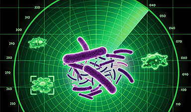
The Secret “Radar” Bacteria Use To Outsmart Their Enemies
A chemical radar allows bacteria to sense and eliminate predators. Investigating how microorganisms communicate deepens our understanding of the complex ecological interactions that shape our environment is an area of key focus for the [...]

Psychologists explore ethical issues associated with human-AI relationships
It's becoming increasingly commonplace for people to develop intimate, long-term relationships with artificial intelligence (AI) technologies. At their extreme, people have "married" their AI companions in non-legally binding ceremonies, and at least two people [...]
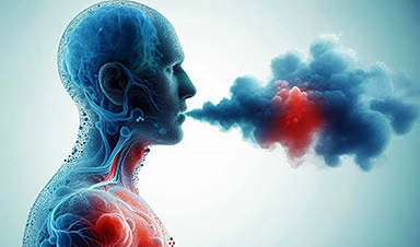
When You Lose Weight, Where Does It Actually Go?
Most health professionals lack a clear understanding of how body fat is lost, often subscribing to misconceptions like fat converting to energy or muscle. The truth is, fat is actually broken down into carbon [...]
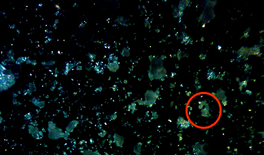
How Everyday Plastics Quietly Turn Into DNA-Damaging Nanoparticles
The same unique structure that makes plastic so versatile also makes it susceptible to breaking down into harmful micro- and nanoscale particles. The world is saturated with trillions of microscopic and nanoscopic plastic particles, some smaller [...]
AI Outperforms Physicians in Real-World Urgent Care Decisions, Study Finds
The study, conducted at the virtual urgent care clinic Cedars-Sinai Connect in LA, compared recommendations given in about 500 visits of adult patients with relatively common symptoms – respiratory, urinary, eye, vaginal and dental. [...]

Challenging the Big Bang: A Multi-Singularity Origin for the Universe
In a study published in the journal Classical and Quantum Gravity, Dr. Richard Lieu, a physics professor at The University of Alabama in Huntsville (UAH), which is a part of The University of Alabama System, suggests that [...]

New drug restores vision by regenerating retinal nerves
Vision is one of the most crucial human senses, yet over 300 million people worldwide are at risk of vision loss due to various retinal diseases. While recent advancements in retinal disease treatments have [...]
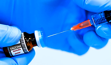
Shingles vaccine cuts dementia risk by 20%, new study shows
A shingles shot may do more than prevent rash — it could help shield the aging brain from dementia, according to a landmark study using real-world data from the UK. A routine vaccine could [...]

AI Predicts Sudden Cardiac Arrest Days Before It Strikes
AI can now predict deadly heart arrhythmias up to two weeks in advance, potentially transforming cardiac care. Artificial intelligence could play a key role in preventing many cases of sudden cardiac death, according to [...]

NanoApps Medical is a Top 20 Feedspot Nanotech Blog
There is an ocean of Nanotechnology news published every day. Feedspot saves us a lot of time and we recommend it. We have been using it since 2018. Feedspot is a freemium online RSS [...]
This Startup Says It Can Clean Your Blood of Microplastics
This is a non-exhaustive list of places microplastics have been found: Mount Everest, the Mariana Trench, Antarctic snow, clouds, plankton, turtles, whales, cattle, birds, tap water, beer, salt, human placentas, semen, breast milk, feces, testicles, [...]

New Blood Test Detects Alzheimer’s and Tracks Its Progression With 92% Accuracy
The new test could help identify which patients are most likely to benefit from new Alzheimer’s drugs. A newly developed blood test for Alzheimer’s disease not only helps confirm the presence of the condition but also [...]
The CDC buried a measles forecast that stressed the need for vaccinations
This story was originally published on ProPublica, a nonprofit newsroom that investigates abuses of power. Sign up to receive our biggest stories as soon as they’re published. ProPublica — Leaders at the Centers for Disease Control and Prevention [...]
Light-Driven Plasmonic Microrobots for Nanoparticle Manipulation
A recent study published in Nature Communications presents a new microrobotic platform designed to improve the precision and versatility of nanoparticle manipulation using light. Led by Jin Qin and colleagues, the research addresses limitations in traditional [...]
Cancer’s “Master Switch” Blocked for Good in Landmark Study
Researchers discovered peptides that permanently block a key cancer protein once thought untreatable, using a new screening method to test their effectiveness inside cells. For the first time, scientists have identified promising drug candidates [...]


