Researchers found that DNA mutations from antiviral enzymes and chemotherapy fuel early bladder cancer, while abnormal circular DNA in tumor cells drives resistance to therapy. These discoveries open new therapeutic avenues.
A groundbreaking study led by researchers at Weill Cornell Medicine and the New York Genome Center has provided unprecedented insights into how bladder cancer begins and progresses. The team discovered that antiviral enzymes, which mutate the DNA of both normal and cancer cells, are key promoters of early bladder cancer development, and that standard chemotherapy is also a potent source of mutations.
The researchers also discovered that overactive genes within abnormal circular DNA structures in tumor cells genes drive bladder cancer resistance to therapy. These findings are novel insights into bladder cancer biology and point to new therapeutic strategies for this difficult-to-treat cancer.
The study, published recently in Nature, focused on the main form of bladder cancer, urothelial carcinoma, which originates from cells that line the bladder, urethra, and tubes that drain urine from the kidneys. The researchers examined malignant and pre-malignant urothelial cells taken from the same set of patients at different disease stages. They used whole-genome sequencing and advanced computational methods to map common DNA mutations, complex structural variants, and their timing.
“Our findings define new fundamental mechanisms driving bladder cancer evolution—mechanisms that we can now think about targeting with therapies,” said co-senior author Dr. Bishoy Faltas, the Gellert Family–John P. Leonard MD Research Scholar in Hematology and Medical Oncology and an associate professor of medicine and of cell and developmental biology at Weill Cornell Medicine, and an oncologist at NewYork-Presbyterian/Weill Cornell Medical Center.
Dr. Nicolas Robine, director of computational biology at the New York Genome Center, and Dr. Olivier Elemento, director of the Englander Institute for Precision Medicine and a professor of physiology and biophysics at Weill Cornell Medicine, also led the study with Dr. Faltas. The co-first authors were Duy Nguyen, a technician in the Faltas Laboratory (now a doctoral student at Harvard Medical School); William Hooper, a bioinformatics scientist at the New York Genome Center; and Dr. Weisi Liu, an instructor in the Faltas Laboratory.
Major Therapeutic Targets Come into Focus
Bladder cancer occurs at the rate of about 80,000 cases per year in the United States. It can be cured with surgery if caught early, but about 30 percent of cases are diagnosed at later stages when it is much harder to treat successfully.
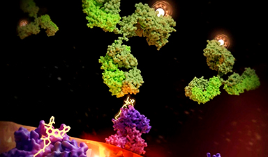
The researchers in the new study found strong evidence that the APOBEC3 enzymes cause early mutations that may help trigger the development of this cancer type. These enzymes evolved to disable infecting retroviruses by editing their viral DNA, though it is known that they can sometimes mutate cells’ own DNA.
“The exact role of APOBEC3-induced mutations in cancer initiation hasn’t been clear,” said Dr. Faltas, who is also the chief research officer at the Englander Institute for Precision Medicine and a member of the Sandra and Edward Meyer Cancer Center at Weill Cornell Medicine. “But we found that these mutations appear early in urothelial cancer, occurring even in pre-malignant urothelial tissue.” In his lab, Dr. Faltas is focusing on studying the role of these mutagenic enzymes in driving cancer evolution.
The researchers found that cisplatin and other platinum-based chemotherapies cause further prominent bursts of mutations, some of which likely allow urothelial cancer cells to survive better and spread despite treatment.
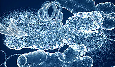
A third major finding was that urothelial tumors often contain complex rearrangements of their DNA that give rise to circular segments of DNA. These “extra-chromosomal DNAs” (ecDNAs) exist apart from chromosomes in the cell nucleus and can sometimes harbor hundreds of copies of cancer-driving growth genes. The researchers discovered that these ecDNA events persist and become more complex, incorporating new DNA segments after treatment, suggesting that they drive resistance to therapy.
This prompted the team to experimentally model an ecDNA version of one of these genes, called CCND1, a master regulator of the cell cycle in the laboratory. The results of these experiments confirmed that CCND1 in this extrachromosomal configuration drives treatment resistance.
Altogether, the findings paint a much clearer picture of the factors that trigger and drive urothelial cancer.
“Traditionally, when analyzing tumor genomes, we’ve used methods that analyze only a tiny fraction of their DNA, but we’ve come to realize that there’s a lot more to discover if we sequence all their DNA and use smart methods to evaluate that data,” Dr. Elemento said. “I think this collaboration vindicates that strategy.”
The Englander Institute and New York Genome Center researchers are planning larger future collaborative studies to dig even deeper into urothelial cancer biology, for example, doing whole-genome sequencing of DNA along with readouts of gene activity not just in bulk tumor samples but in individual tumor cells.
“Combining those two sets of information at the single-cell level would be tremendously important and interesting,” Dr. Robine said.
The researchers also plan to study potential clinical applications of this work. The investigators are hopeful that a new FDA-approved drug targeting the ERBB2 gene product—the HER2 receptor protein, also found on breast tumor cells—will work especially well in urothelial cancer patients with strong signs of ERBB2 ecDNAs. They are also working on finding ways to block ecDNA formation and maintenance.
Reference: “The interplay of mutagenesis and ecDNA shapes urothelial cancer evolution” by Duy D. Nguyen, William F. Hooper, Weisi Liu, Timothy R. Chu, Heather Geiger, Jennifer M. Shelton, Minita Shah, Zoe R. Goldstein, Lara Winterkorn, Adrienne Helland, Michael Sigouros, Jyothi Manohar, Jenna Moyer, Majd Al Assaad, Alissa Semaan, Sandra Cohen, Florencia Madorsky Rowdo, David Wilkes, Mohamed Osman, Rahul R. Singh, Andrea Sboner, Henkel L. Valentine, Phillip Abbosh, Scott T. Tagawa, David M. Nanus, Jones T. Nauseef, Cora N. Sternberg, Ana M. Molina, Douglas Scherr, Giorgio Inghirami, Juan Miguel Mosquera, Olivier Elemento, Nicolas Robine and Bishoy M. Faltas, 9 October 2024, Nature.
DOI: 10.1038/s41586-024-07955-3
The research reported in this story was supported in part by the National Cancer Institute and the National Center for Advancing Translational Science, both part of the National Institutes of Health, through grant numbers R37CA279737, U01CA260369, UL1TR002384; and the United States Department of Defense through grant number W81XWH-17-1-0539. Additional support was provided by the Starr Cancer Consortium, the Leo & Anne Albert Institute for Bladder Cancer Care and Research, the Translational Research Program in the Department of Pathology and Laboratory Medicine at Weill Cornell Medicine, and the New York Genome Center’s Polyethnic-1000 Initiative.
News
New mpox variant can spread rapidly across borders
International researchers, including from DTU National Food Institute, warn that the ongoing mpox outbreak in the Democratic Republic of the Congo (DRC) has the potential to spread across borders more rapidly. The mpox virus [...]
How far would you trust AI to make important decisions?
From tailored Netflix recommendations to personalized Facebook feeds, artificial intelligence (AI) adeptly serves content that matches our preferences and past behaviors. But while a restaurant tip or two is handy, how comfortable would you [...]
Can AI Really Think? Research Reveals Gaps in Logical Execution
While AI models can break down problems into structured steps, new research reveals they still fail at basic arithmetic and fact-checking—raising questions about their true reasoning abilities. Large Language Models (LLMs) have become indispensable [...]
Scientists Just Made Cancer Radiation Therapy Smarter, Safer, and More Precise
Scientists at UC San Francisco have developed a revolutionary cancer treatment that precisely targets tumors with radiation while sparing healthy tissues. By using a KRAS-targeting drug to mark cancer cells and attaching a radioactive [...]
Superbugs Are Losing to Science, Light, and a Little Spice
Texas A&M researchers have found that curcumin, when activated by light, can weaken antibiotic-resistant bacteria, restoring the effectiveness of conventional antibiotics. Curcumin: A Surprising Ally Against Superbugs In 2017, a woman admitted to a [...]
New Research Shatters the Perfect Pitch Myth
For decades, people believed absolute pitch was an exclusive ability granted only to those with the right genetics or early music training. But new research from the University of Surrey proves otherwise. It’s been [...]
Why Some Drinkers Suffer Devastating Liver Damage While Others Don’t
A study from Keck Medicine of USC found that heavy drinkers with diabetes, high blood pressure, or a large waistline are up to 2.4 times more likely to develop advanced liver disease. These conditions may amplify [...]
“Good” Cholesterol Could Be Bad for Your Eyes – New Study Raises Concerns
‘Good’ cholesterol may be linked to an increased risk of glaucoma in individuals over 55, while, paradoxically, ‘bad’ cholesterol may be associated with a lower risk. These findings challenge conventional beliefs about factors that [...]
Reawakening Dormant Nerve Cells: Groundbreaking Neurotechnology Restores Motor Function
A new electrical stimulation therapy for spinal muscle atrophy (SMA) has shown promise in reactivating motor neurons and improving movement. In a pilot clinical trial, three patients who received spinal cord stimulation for one [...]
AI’s Energy Crisis Solved? A Revolutionary Magnetic Chip Could Change Everything
AI is evolving at an incredible pace, but its growing energy demands pose a major challenge. Enter spintronic devices—new technology that mimics the brain’s efficiency by integrating memory and processing. Scientists in Japan have [...]
Nanotechnology for oil spill response and cleanup in coastal regions
(Nanowerk News) Cleaning up after a major oil spill is a long, expensive process, and the damage to a coastal region’s ecosystem can be significant. This is especially true for the world’s Arctic region, [...]
The Role of Nanotechnology in Space Exploration
Nanotechnology, which involves working with materials at the atomic or molecular level, is becoming increasingly important in space exploration. By improving strength, thermal stability, electrical conductivity, and radiation resistance, nanotechnology is helping create lighter, more [...]
New Study Challenges Beliefs About CBD in Pregnancy, Reveals Unexpected Risks
CBD is gaining popularity as a remedy for pregnancy symptoms like nausea and anxiety, but new research suggests it may not be as safe as many believe. A study from McMaster University found that [...]
Does COVID increase the risk of Alzheimer’s disease?
Scientists discover that even mild COVID-19 can alter brain proteins linked to Alzheimer’s disease, potentially increasing dementia risk—raising urgent public health concerns. A recent study published in the journal Nature Medicine investigated whether both mild and [...]
New MRI Study Reveals How Cannabis Alters Brain Activity and Weakens Memory
A massive new study sheds light on how cannabis affects the brain, particularly during cognitive tasks. Researchers analyzed over 1,000 young adults and found that both heavy lifetime use and recent cannabis consumption significantly reduced brain [...]
How to Assess Nanotoxicity: Key Methods and Protocols
With their high surface area and enhanced physicochemical properties, nanomaterials play a critical role in drug delivery, consumer products, and environmental technologies. However, their nanoscale dimensions enable interactions with cellular components in complex and [...]
