A cutting-edge X-ray method reveals the 3D orientation of nanoscale material structures, offering fresh insights into their functionality.
Researchers at the Swiss Light Source (SLS) have developed a groundbreaking technique called X-ray linear dichroic orientation tomography (XL-DOT). This method reveals the three-dimensional arrangement of a material's structural building blocks at the nanoscale. Its first application focused on a polycrystalline catalyst, enabling scientists to visualize crystal grains, grain boundaries, and defects—critical features that influence catalyst performance. Beyond catalysis, XL-DOT offers unprecedented insights into the structure of various functional materials used in information technology, energy storage, and biomedical applications.
Advancements in Non-Destructive Imaging of Material Microstructures
Zooming into the micro- or nanostructure of functional materials — whether natural or man-made — reveals countless coherent domains or grains. These grains are distinct regions where molecules and atoms are arranged in orderly, repeating patterns.
The arrangement of these grains is closely tied to the material's properties. Their size, orientation, and distribution can mean the difference between a sturdy brick and a crumbling stone. They determine how ductile a metal is, how efficiently a semiconductor transfers electrons, and how well ceramics conduct heat. This structural organization also plays a critical role in biological materials; for example, collagen fibers are made of interwoven fibrils, and their alignment affects the mechanical strength of connective tissues.
These domains are often tiny: tens of nanometers in size. And it is their arrangement in three dimensions over extended volumes that is property-determining. Yet until now, techniques to probe the organization of materials at the nanoscale have largely been confined to two dimensions or are destructive in nature.
Now, using X-rays generated by the Swiss Light Source SLS, a collaborative team of researchers from Paul Scherrer Institute PSI, ETH Zurich, the University of Oxford and the Max Plank Institute for Chemical Physics of Solids have succeeded in creating an imaging technique to access this information in three-dimensions.
"We Not Only Look Inside, but With Nanoscale Resolution"
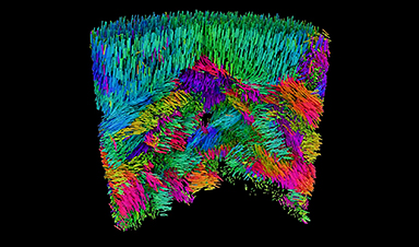
Their technique is known as X-ray linear dichroic orientation tomography, or XL-DOT for short. XL-DOT uses polarized X-rays from the Swiss Light Source SLS, to probe how materials absorb X-rays differently depending on the orientation of structural domains inside. By changing the polarization of the X-rays, while rotating the sample to capture images from different angles, the technique creates a three-dimensional map revealing the internal organization of the material.
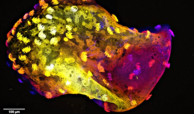
The team applied their method to a chunk of vanadium pentoxide catalyst about one micron in diameter, used in the production of sulfuric acid. Here, they could identify minute details in the catalyst`s structure including crystalline grains, boundaries where grains meet, and changes in the crystal orientation. They also identified topological defects in the catalyst. Such features directly affect the activity and stability of catalysts, so knowledge of this structure is crucial in optimizing performance.
Importantly, the method achieves high spatial resolution. Because X-rays have a short wavelength, the method can resolve structures just tens of nanometers in size, aligning with the sizes of features such as the crystalline grains.
"Linear dichroism has been used to measure anisotropies in materials for many years, but this is the first time it has been extended to 3D. We not only look inside, but with nanoscale resolution," says Valerio Scagnoli, Senior Scientist in the Mesoscopic Systems, a joint group between PSI and ETH Zurich. "This means that we now have access to information that was not previously visible, and we can achieve this in small but representative samples, several micrometers in size."
Leading the way with coherent X-rays
Although the researchers first had the idea for XL-DOT in 2019, it would take another five years to put it into practice. Together with complex experimental requirements, a major hurdle was extracting the three-dimensional map of crystal orientations from terabytes of raw data. This mathematical puzzle was overcome with the development of a dedicated reconstruction algorithm by Andreas Apseros, first author of the study, during his doctoral studies at PSI, funded by the Swiss National Science Foundation (SNSF).
The researchers believe that their success in developing XL-DOT is in part thanks to the long-term commitment to developing expertise with coherent X-rays at PSI, which led to unprecedented control and instrument stability at the coherent Small Angle X-ray Scattering (cSAXS) beamline: critical for the delicate measurements.
This is an area that is set to leap forward after the SLS 2.0 upgrade: "Coherence is where we're really set to gain with the upgrade," says Apseros. "We're looking at very weak signals, so with more coherent photons, we'll have more signal and can either go to more difficult materials or higher spatial resolution."
A way into the microstructure of diverse materials
Given the non-destructive nature of XL-DOT, the researchers foresee operando investigations of systems such as batteries as well as catalysts. "Catalyst bodies and cathode particles in batteries are typically between ten and fifty micrometers in size, so this is a reasonable next step," says Johannes Ihli, formerly of cSAXS and currently at the University of Oxford, who led the study.
Yet the new technique is not just useful for catalysts, the researchers emphasize. It is useful for all types of materials that exhibit ordered microstructures, whether biological tissues or advanced materials for information technology or energy storage.
Indeed, for the research team, the scientific motivation lies with probing the three-dimensional magnetic organization of materials. An example is the orientation of magnetic moments within antiferromagnetic materials. Here, the magnetic moments are aligned in alternating directions when going from atom to atom. Such materials maintain no net magnetization when measured at a distance, yet they do possess local order in the magnetic structure, a fact that is appealing for technological applications such as faster and more efficient data processing. "Our method is one of the only ways to probe this orientation," says Claire Donnelly, group leader Max Planck Institute for Chemical Physics of Solids in Dresden who, since carrying out her doctoral work in the Mesoscopic Systems group has maintained a strong collaboration with the team at PSI.
It was during this doctoral work that Donnelly together with the same team at PSI published in Nature a method to carry out magnetic tomography using circularly polarized X-rays (in contrast to XL-DOT, which uses linearly polarized X-rays). This has since been implemented in synchrotrons around the world.
With the groundwork for XL-DOT laid, the team hope that it will, in a similar way to its circularly polarized sibling, become a widely used technique at synchrotrons. Given the much wider range of samples that XL-DOT is relevant to and the importance of structural ordering to material performance, the impact of this latest method may be expected to be even greater. "Now that we've overcome many of the challenges, other beamlines can implement the technique. And we can help them to do it," adds Donnelly.
Reference: "X-ray Linear Dichroic Tomography of Crystallographic and Topological Defects" 11 December 2024, Nature.
News

Scientists Uncover Fatal Weakness in “Zombie Cells” Linked to Cancer
A newly identified weakness in “zombie” cells may open the door to more precise cancer treatments by turning their own survival strategy against them. A new class of drugs takes advantage of a recently [...]

Bowel and Ovarian Cancers Are Dramatically Rising in Young Adults, Scientists Aren’t Sure Why
Cancer incidence is increasing, especially among younger adults, and current risk factors don’t fully account for the trend. Scientists suggest other underlying causes may be contributing. Cancer patterns in England are shifting in a [...]

New Immune Pathway Could Supercharge mRNA Cancer Vaccines
A surprising backup system in the immune response to mRNA vaccines may hold the key to more effective cancer treatments. The arrival of mRNA vaccines against SARS-CoV-2 in 2020 marked a turning point in the COVID-19 pandemic. Today, [...]

Scientists Discover “Molecular Switch” That Fuels Alzheimer’s Brain Inflammation
A newly identified trigger of brain inflammation could offer a fresh target for slowing Alzheimer’s progression. The brain has its own built-in immune system that identifies threats and responds to them. In Alzheimer’s disease, growing evidence [...]

Molecular Manufacturing: The Future of Nanomedicine – New book from NanoappsMedical Inc.
This book explores the revolutionary potential of atomically precise manufacturing technologies to transform global healthcare, as well as practically every other sector across society. This forward-thinking volume examines how envisaged Factory@Home systems might enable the cost-effective [...]

Forgotten Medicinal Plant Shows Promise in Fighting Dangerous Superbugs
A traditional medicinal plant, tormentil, shows promise against antibiotic-resistant bacteria in laboratory tests. Its compounds work by limiting bacterial growth and boosting antibiotic performance. Before the development of modern antibiotics, plant-based remedies were commonly [...]

NanoMedical Brain/Cloud Interface – Explorations and Implications. A new book from Frank Boehm
New book from Frank Boehm, NanoappsMedical Inc Founder: This book explores the future hypothetical possibility that the cerebral cortex of the human brain might be seamlessly, safely, and securely connected with the Cloud via [...]

New Research Finds Shocking Link Between Chili Peppers and Cancer
If you love spicy food, you are not alone. But scientists are taking a closer look at whether eating a lot of chili peppers could affect your cancer risk. Could your love of spicy [...]

New book from Nanoappsmedical Inc. – Global Health Care Equivalency
A new book by Frank Boehm, NanoappsMedical Inc. Founder. This groundbreaking volume explores the vision of a Global Health Care Equivalency (GHCE) system powered by artificial intelligence and quantum computing technologies, operating on secure [...]
Scientists Create “Neurobots” – Living Machines With Their Own Nervous Systems
Neurobots—xenobots with neurons—show self-organized nervous systems and enhanced behaviors, revealing new insights into how biology builds functional structures. In 2020, researchers at Tufts University developed tiny living structures known as xenobots using frog cells. These microscopic organisms [...]

Our books now available worldwide!
Online Sellers other than Amazon, Routledge, and IOPP Indigo Global Health Care Equivalency in the Age of Nanotechnology, Nanomedicine and Artifcial Intelligence Global Health Care Equivalency In The Age Of Nanotechnology, Nanomedicine And Artificial [...]

Amazonian Chocolate Could Become the Next Superfood, Scientists Say
New research into Amazonian cocoa reveals that its value may extend beyond flavor alone. Chocolate from the Amazon is already known worldwide for its distinctive taste, but new research suggests it may offer even [...]

Nanobody repairs misfolded CFTR inside cells, boosting function in cystic fibrosis
A tiny antibody component could fundamentally transform the treatment of cystic fibrosis: For the first time, researchers have succeeded in developing a so-called nanobody that penetrates directly into human cells and can repair the [...]

20-Year Study Finds Daily Multivitamins Don’t Extend Lifespan
A large, decades-long study of over 390,000 U.S. adults challenges a widespread assumption about daily multivitamins. Multivitamins are a daily habit for millions of Americans, often taken with the expectation that they will extend [...]

Novel Investment Paradigms for Regenerative Healthcare Ecosystems
Introduction The transition toward regenerative healthcare ecosystems—anchored in wellness optimization, disease prevention, eradication strategies, and healthy longevity—necessitates a structural reconfiguration of capital architectures, governance models, and incentive design. Regenerative healthcare, by definition, transcends episodic [...]

What If Consciousness Exists Beyond Your Brain
Scientists still don’t know how consciousness emerges from the brain. New ideas suggest it may not emerge at all, but instead be a basic feature of reality. Is consciousness produced by the brain, or [...]