
Hyperspectral microscopy is an advanced visualization technique that combines hyperspectral imaging with state-of-the-art optics and computer software to enable rapid identification of nanomaterials. Since hyperspectral datacubes are large, their acquisition is complicated and time-consuming.
Despite the efficiency of spectral scanning in acquiring hyperspectral datacubes, this technique cannot be extended to large numbers of spectral bands because of their low light levels due to narrowband filters and mechanical difficulties while using large filter wheels.
However, applying a digital micromirror device (DMD) can circumvent the above drawbacks during spectral multiplexing. Utilizing a single DMD avoids the need for large filter wheels by promoting arbitrary spectral programming.
In an article published in The Journal of Physical Chemistry C, a brightfield DMD-based multiplexing microscope was employed to investigate the two-dimensional (2D) nanomaterials. Furthermore, the effectiveness of the DMD-based microscopy was demonstrated by measuring the thickness of few-layer graphene and molybdenum sulfide (MoS2) from their corresponding contrast spectra, which were later compared to their theoretical curves for validation.
Hyperspectral Microscopy to Characterize 2D Materials
Atomically thin semiconducting 2D materials are extensively applied in nanophotonics, and the outstanding optical properties of these 2D materials play a critical role in many applications. Hence, accurate characterization of these 2D materials is critical to employ them in device structures to pattern the necessary electrical contacts.
Hyperspectral microscopy is a spectral imaging modality that can obtain a sample’s full spectroscopic information and render it in image form, and is one technique that is being developed and explored to address current analytical challenges for nanoscale 2D materials.
Hyperspectral microscopy involves the functional combination of a traditional high-resolution microscope and spectrometer. The motivation behind developing this technique for biomedical applications comes from an interest in the biological sample’s emission or reflectance spectrum, which contains important structural, biochemical, or physiological information.
The unique optical properties of 2D materials are largely dependent on the number of atomic layers. Hyperspectral imaging microscopy shows a large potential for rapid and accurate thickness mapping.
Hyperspectral Microscopy of 2D Materials
In the present study, the DMD was employed to encode the illumination source’s spectral content and overcome the mechanical difficulties of hyperspectral microscopy in terms of imaging with a filter wheel. This method promoted Hadamard multiplexing in the spectral content of the sample, improving the light throughput without affecting the signal-to-noise ratio.
Although using DMD as a programmable spectral filter was previously reported, this was the first work that applied it to hyperspectral microscopy of nanomaterials. The proposed multiplexing microscope was composed of illumination and an imager. While the illumination side was employed with a hyperspectral projector, the imager consisted of a reflective brightfield microscope.
Moreover, the microscope’s entrance had a biconvex lens that focused the incident light to the back focal plane of the objective to realize Koehler’s illumination. On the other hand, the objective lens focused the illumination that was spectrally programmed down to the sample and collected the light reflected.
The bandwidth and spectral resolution of the microscope were measured using tantalum sulfide (TaS2) since it is highly reflective across the visible region. The two hyperspectral images obtained revealed that the topographical features in transmission mode were more than in reflection mode.
Measuring the exciton peaks in MoS2 and comparing them to the theoretical result computed using Fresnel’s equations showed good agreement with the theoretical spectra for monolayer and bilayer MoS2.
Furthermore, the image of graphite nanosheets at the camera and the reconstructed hyperspectral image showed regions with multiple spatially separated flakes. The reconstructed image helped to optically determine the thickness of the flakes at different parts of the nanosheet.
Conclusion
In summary, diffraction-limited, fast, large-field-of-view hyperspectral microscopy was demonstrated to contrast spectroscopy. The proposed system could be applied for characterizing novel devices and thin film heterostructures. Additional modifications to the hyperspectral microscope can enable different experiments.
For example, the sample, transmission, and reflection hyperspectral imaging can be concurrently achieved with a long working distance objective. Hyperspectral imaging of TaS2 with three regions of differing thickness revealed that the topographical features in transmission mode were more than in reflection mode.
On the other hand, for the samples that evolve over time, performing hyperspectral video microscopy allowed sampling of both spectral and temporal dimensions. Moreover, single-pixel imaging could be naturally incorporated into the system by utilizing DMD and a single detector instead of a camera.
This enabled hyperspectral microscopy in the infrared, which otherwise becomes expensive for cameras. The spatial, temporal, and spectral information was captured on a single detector followed by reconstruction using compressive sensing recovery algorithms.
News

Scientists Invent Plastic That Can Dissolve In Seawater In Just A Few Hours
Plastic waste and pollution in the sea have been among the most serious environmental problems for decades, causing immense damage to marine life and ecosystems. However, a breakthrough discovery may offer a game-changing solution. [...]
Muscles from the 3D printer
Swiss researchers have developed a method for printing artificial muscles out of silicone. In the future, these could be used on both humans and robots. Swiss researchers have succeeded in printing artificial muscles out [...]
Beneficial genetic changes observed in regular blood donors
Researchers at the Francis Crick Institute have identified genetic changes in blood stem cells from frequent blood donors that support the production of new, non-cancerous cells. Understanding the differences in the mutations that accumulate [...]

Shocking Amounts of Microplastics in the Brain – It Could Be Increasing Our Risk of Dementia
The brain has higher concentrations of plastic particles compared to other organs, with increased levels found in dementia patients. In a comprehensive commentary published in Brain Medicine, researchers highlight alarming new evidence of microplastic accumulation [...]

Baffling Scientists for Centuries: New Study Unravels Mystery of Static Electricity
ISTA physicists demonstrate that contact electrification depends on the contact history of materials. For centuries, static electricity has intrigued and perplexed scientists. Now, researchers from the Waitukaitis group at the Institute of Science and [...]

Tumor “Stickiness” – Scientists Develop Potential New Way To Predict Cancer’s Spread
UC San Diego researchers have developed a device that predicts breast cancer aggressiveness by measuring tumor cell adhesion. Weakly adherent cells indicate a higher risk of metastasis, especially in early-stage DCIS. This innovation could [...]

Scientists Just Watched Atoms Move for the First Time Using AI
Scientists have developed a groundbreaking AI-driven technique that reveals the hidden movements of nanoparticles, essential in materials science, pharmaceuticals, and electronics. By integrating artificial intelligence with electron microscopy, researchers can now visualize atomic-level changes that were [...]

Scientists Sound Alarm: “Safe” Antibiotic Has Led to an Almost Untreatable Superbug
A recent study reveals that an antibiotic used for liver disease patients may increase their risk of contracting a dangerous superbug. An international team of researchers has discovered that rifaximin, a commonly prescribed antibiotic [...]
Scientists Discover Natural Compound That Stops Cancer Progression
A discovery led by OHSU was made possible by years of study conducted by University of Portland undergraduates. Scientists have discovered a natural compound that can halt a key process involved in the progression [...]

Scientists Just Discovered an RNA That Repairs DNA Damage – And It’s a Game-Changer
Our DNA is constantly under threat — from cell division errors to external factors like sunlight and smoking. Fortunately, cells have intricate repair mechanisms to counteract this damage. Scientists have uncovered a surprising role played by [...]

What Scientists Just Discovered About COVID-19’s Hidden Death Toll
COVID-19 didn’t just claim lives directly—it reshaped mortality patterns worldwide. A major international study found that life expectancy plummeted across most of the 24 analyzed countries, with additional deaths from cardiovascular disease, substance abuse, and mental [...]
Self-Propelled Nanoparticles Improve Immunotherapy for Non-Invasive Bladder Cancer
A study led by Pohang University of Science and Technology (POSTECH) and the Institute for Bioengineering of Catalonia (IBEC) in South Korea details the creation of urea-powered nanomotors that enhance immunotherapy for bladder cancer. The nanomotors [...]
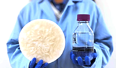
Scientists Develop New System That Produces Drinking Water From Thin Air
UT Austin researchers have developed a biodegradable, biomass-based hydrogel that efficiently extracts drinkable water from the air, offering a scalable, sustainable solution for water access in off-grid communities, emergency relief, and agriculture. Discarded food [...]

AI Unveils Hidden Nanoparticles – A Breakthrough in Early Disease Detection
Deep Nanometry (DNM) is an innovative technique combining high-speed optical detection with AI-driven noise reduction, allowing researchers to find rare nanoparticles like extracellular vesicles (EVs). Since EVs play a role in disease detection, DNM [...]
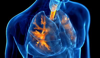
Inhalable nanoparticles could help treat chronic lung disease
Nanoparticles designed to release antibiotics deep inside the lungs reduced inflammation and improved lung function in mice with symptoms of chronic obstructive pulmonary disease By Grace Wade Delivering medication to the lungs with inhalable nanoparticles [...]
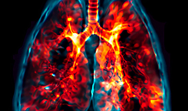
New MRI Study Uncovers Hidden Lung Abnormalities in Children With Long COVID
Long COVID is more than just lingering symptoms—it may have a hidden biological basis that standard medical tests fail to detect. A groundbreaking study using advanced MRI technology has uncovered significant lung abnormalities in [...]


