
Long COVID is more than just lingering symptoms—it may have a hidden biological basis that standard medical tests fail to detect.
A groundbreaking study using advanced MRI technology has uncovered significant lung abnormalities in children and adolescents suffering from long COVID, particularly in blood flow and air movement. These findings help explain persistent symptoms like chronic fatigue and shortness of breath, offering a new path for diagnosing and managing this condition.
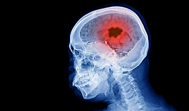
MRI Reveals Lung Abnormalities in Children with Long COVID
A new study published today (February 25) in Radiology, the journal of the Radiological Society of North America (RSNA), reveals that an advanced type of MRI has detected significant lung abnormalities in children and adolescents with long COVID.
Long COVID, or post-COVID-19 condition, occurs when symptoms persist for more than 12 weeks after a COVID-19 infection. While children and teens generally experience a milder form of the condition, symptoms like chronic fatigue, headaches, and difficulty concentrating can interfere with school and social activities.
A Need for Better Testing
In adults, chest CT scans are commonly used to assess lung function in long COVID cases. However, this method is not usually recommended for children due to radiation exposure and the potential need for contrast agents.
Instead, young patients suspected of having long COVID are typically evaluated through pulmonary function tests, echocardiography, and medical history reviews. Unfortunately, these standard tests often show normal lung and heart function, even in children experiencing ongoing symptoms.
"Parents should understand that their children's persistent symptoms after COVID-19 may have a measurable physiological basis, even when standard medical tests appear normal," said lead study author Gesa H. Pöhler, M.D., a senior physician in the Department of Diagnostic and Interventional Radiology at Hannover Medical School in Germany.
The researchers employed phase-resolved functional lung (PREFUL) MRI. This advanced MRI technology can analyze lung ventilation (air movement in and out of the lungs) and perfusion (blood flow through the lungs). PREFUL MRI doesn't require the use of radiation or intravenous contrast agents and can be done while the patient breathes freely, making it a suitable procedure for children.
First Evidence of Lung Perfusion Abnormalities
"Our research provides the first comprehensive evidence of measurable regional lung perfusion abnormalities in pediatric post-COVID-19 condition using radiation-free, contrast-free lung imaging," Dr. Pöhler said.
For the prospective study, conducted between April 2022 and 2023, the researchers enrolled 54 patients ranging in age from 11 to 17 years. Half of the patients were diagnosed with long COVID, and the other half were healthy controls. A self-reported assessment called the bell score was used to assess symptom severity in patients with long COVID.

Blood Flow Reduction and Fatigue Connection
Compared to healthy controls, children and adolescents with long COVID had significantly reduced blood flow in the lungs. A reduction in blood flow patterns in organs or other areas of the body can result in a lack of sufficient oxygen and nutrients.
The most prevalent symptom of fatigue affected all but one patient with long COVID.
"Importantly, the severity of fatigue symptoms correlated with these blood flow changes, suggesting a possible biological basis for the patients' ongoing symptoms," Dr. Pöhler said.
In addition to poor blood flow, a subgroup of long COVID patients with cardiopulmonary symptoms, such as shortness of breath, also showed a reduction of air movement and reach in the lungs.

Future Implications for Long COVID Monitoring
The researchers suggest that continuous monitoring of lung abnormalities in children with long COVID at various stages of the condition could help guide therapeutic interventions and monitoring strategies.
"Quantitative lung MRI establishes a potential imaging biomarker profiling and helps to enable disease severity follow-up for this complex condition in the future," Dr. Pöhler said.
Reference: "Phase-resolved Functional Lung MRI Reveals Distinct Lung Perfusion Phenotype in Children and Adolescents with Post–COVID-19 Condition" by Gesa H. Pöhler, Andreas Voskrebenzev, Marc-Luca Heinze, Valentina Skeries, Filip Klimeš, Julian Glandorf, Jan Eckstein, Nigar Babazade, Marius Wernz, Alexander Pfeil, Gesine Hansen, Frank K. Wacker, Jens Vogel-Claussen, Martin Wetzke and Diane Miriam Renz, 25 February 2025, Radiology.
DOI: 10.1148/radiol.241596
Collaborating with Dr. Pöhler were Andreas Voskrebenzev, Ph.D., Marc-Luca Heinze, Valentina Skeries, M.D., Filip Klimeš, Ph.D., Julian Glandorf, M.D., Jan Eckstein, M.D., Nigar Babazade, Marius Wernz, B.S., Alexander Pfeil, M.D., Gesine Hansen, M.D., Frank K. Wacker, M.D., Jens Vogel-Claussen, M.D., Martin Wetzke, M.D., and Diane Miriam Renz, M.D.
News

Scientists Have Discovered These Deadly Parasites Are Secretly Swapping DNA
Leishmania parasites appear to evolve through widespread genetic exchange, reshaping assumptions about how they adapt and spread. A parasite long thought to spread mostly by cloning itself may be far more genetically dynamic than [...]

Stanford’s Revolutionary New Microscope Reveals Living Cells in Stunning Detail
Stanford researchers have developed a microscope that can show how nanostructures interact inside living cells at the highest resolution achieved so far. The view into living cells just got better. Stanford researchers have merged [...]

What Bundibugyo Ebola vaccines and treatments are under development
By Mariam Sunny and Jennifer Rigby May 29 (Reuters) – Global health authorities are racing to identify medical options to help contain an Ebola outbreak in eastern Democratic Republic of Congo, linked to the [...]

Why More People in Their 30s Are Suddenly Getting Colon Cancer
A major Swiss study found that colorectal cancer is becoming increasingly common in adults under 50, even as rates decline in older age groups. Researchers in Switzerland have identified a concerning trend: while colorectal [...]

Researchers Compare MS Models to Human Tissue in Search for Better Therapies
Researchers identified key differences between two widely used multiple sclerosis models, showing how each can better study myelin damage, immune responses, and repair. The findings may improve efforts to develop treatments that restore lost [...]

Scientists Discover Genetic “Off Switch” That Supercharges CAR T Cells Against Cancer
A new study reveals a possible way to make CAR T-cell therapy more durable and effective by targeting a single gene-regulating protein. CAR T-cell therapy is widely seen as a breakthrough in personalized cancer [...]
New Vitamin B12-Based Therapy Could Change How Brain Cancer Is Treated
Researchers have identified a vitamin B12–based compound that appears capable of crossing the blood–brain barrier and selectively accumulating in glioblastoma tissue. For decades, one of the biggest problems in brain cancer treatment has had [...]

Simple Fiber Supplement Cuts Knee Arthritis Pain in Just 6 Weeks, Study Finds
A daily inulin supplement may help reduce knee osteoarthritis pain while revealing a possible link between gut health, muscle function, and pain sensitivity. For millions of people living with knee osteoarthritis, managing chronic pain [...]

This Common Vitamin May Help Stop Prediabetes From Turning Into Diabetes
Vitamin D may help prevent type 2 diabetes in people with specific genetic variations, offering a possible path toward personalized diabetes prevention. More than 40% of U.S. adults have prediabetes, a condition in which [...]

Ebola, hantavirus: Is the world prepared for the next pandemic?
Funding cuts to health research and a growing antivaccine movement are making it harder than ever to respond to viruses. The World Health Organization (WHO) has declared that an Ebola outbreak in Uganda and [...]

May 2026 Healthcare News and Trends: Market Signals That Matter
Artificial intelligence is dominating headlines, telehealth has settled into a new normal, and digital health continues to promise transformation. However, much of what is being discussed in healthcare today reflects potential rather than reality. [...]

Scientists Rewire Donor Stem Cells To Outsmart Aggressive Blood Cancers
Researchers have tested a gene-edited stem cell transplant designed to shield healthy blood-forming cells from powerful cancer-targeting immunotherapies. For patients with highly aggressive blood cancers, stem cell transplantation can offer a rare chance at [...]

Recent Digital Health Trends, Insights and News – May 2026
Last month marked continued progress as digital health moves into its next phase — from AI expanding into drug discovery and core infrastructure to new federal pathways accelerating device access and home-based care. Together, [...]

Cancer Mystery Solved: Scientists Discover How Melanoma Becomes “Immortal”
Scientists have uncovered a previously overlooked mechanism that may help melanoma cells become effectively “immortal.” Cancer cells face a major problem before they can become deadly: They have to figure out how to stop [...]

How Visual Neurons Organize Thousands of Synaptic Inputs
Summary: A new study uncovered the organizational rules that determine how neurons in the primary visual cortex process information. By imaging both the cell bodies (soma) and the individual synapses (on dendritic spines) of [...]

Scientists Just Found a Surprising Way To Destroy “Forever Chemicals”
Scientists have uncovered a new mechanism that may help break down highly persistent PFAS pollutants. PFAS have earned the nickname “forever chemicals” for a reason. These industrial compounds are so chemically durable that they [...]