By Dr Zara Kassam (Drug Target Review):
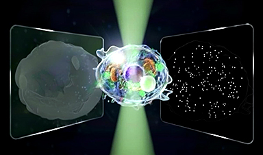
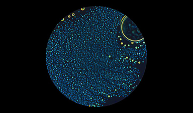
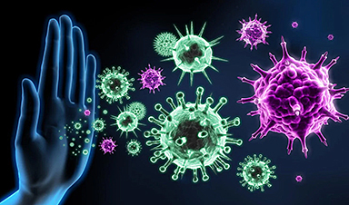
A team of scientists have used a novel live-cell fluorescent imaging system that has allowed them for the first time to identify individual particles associated with HIV.
The researchers at Northwestern Medicine were able to monitor how the HIV capsid uncoats in the cell at the individual particle level. They demonstrated that uncoating leading to infection occurs early in the cytoplasm, around 30 minutes after cell fusion. The finding is just one example of novel discoveries about HIV that might now be possible with the imaging system.
“Being able to connect infectivity of individual particles and how they behave in the cell to infection — which is what we really care about — is going to have a big impact on the field,” said principal investigator Dr Thomas Hope, Professor of Cell and Molecular Biology “The system can now be used to resolve other controversies in HIV biology and to determine which potential targets for drug development are most relevant.”
Recent News
Fake Science Is Growing Faster Than Legitimate Research, New Study Warns
New research reveals organized networks linking paper mills, intermediaries, and compromised academic journals Organized scientific fraud is becoming increasingly common, ranging from fabricated research to the buying and selling [...]
Scientists Unlock a New Way to Hear the Brain’s Hidden Language
Scientists can finally hear the brain’s quietest messages—unlocking the hidden code behind how neurons think, decide, and remember. Scientists have created a new protein that can capture the incoming [...]
Does being infected or vaccinated first influence COVID-19 immunity?
A new study analyzing the immune response to COVID-19 in a Catalan cohort of health workers sheds light on an important question: does it matter whether a person was [...]
We May Never Know if AI Is Conscious, Says Cambridge Philosopher
As claims about conscious AI grow louder, a Cambridge philosopher argues that we lack the evidence to know whether machines can truly be conscious, let alone morally significant. [...]
AI Helped Scientists Stop a Virus With One Tiny Change
Using AI, researchers identified one tiny molecular interaction that viruses need to infect cells. Disrupting it stopped the virus before infection could begin. Washington State University scientists have uncovered a method [...]
Deadly Hospital Fungus May Finally Have a Weakness
A deadly, drug-resistant hospital fungus may finally have a weakness—and scientists think they’ve found it. Researchers have identified a genetic process that could open the door to new treatments [...]
Fever-Proof Bird Flu Variant Could Fuel the Next Pandemic
Bird flu viruses present a significant risk to humans because they can continue replicating at temperatures higher than a typical fever. Fever is one of the body’s main tools [...]
What could the future of nanoscience look like?
Society has a lot to thank for nanoscience. From improved health monitoring to reducing the size of electronics, scientists’ ability to delve deeper and better understand chemistry at the [...]
Scientists Melt Cancer’s Hidden “Power Hubs” and Stop Tumor Growth
Researchers discovered that in a rare kidney cancer, RNA builds droplet-like hubs that act as growth control centers inside tumor cells. By engineering a molecular switch to dissolve these hubs, they [...]
Platelet-inspired nanoparticles could improve treatment of inflammatory diseases
Scientists have developed platelet-inspired nanoparticles that deliver anti-inflammatory drugs directly to brain-computer interface implants, doubling their effectiveness. Scientists have found a way to improve the performance of brain-computer interface [...]
After 150 years, a new chapter in cancer therapy is finally beginning
For decades, researchers have been looking for ways to destroy cancer cells in a targeted manner without further weakening the body. But for many patients whose immune system is severely [...]
Older chemical libraries show promise for fighting resistant strains of COVID-19 virus
SARS‑CoV‑2, the virus that causes COVID-19, continues to mutate, with some newer strains becoming less responsive to current antiviral treatments like Paxlovid. Now, University of California San Diego scientists [...]
Lower doses of immunotherapy for skin cancer give better results, study suggests
According to a new study, lower doses of approved immunotherapy for malignant melanoma can give better results against tumors, while reducing side effects. This is reported by researchers at Karolinska Institutet [...]
Researchers highlight five pathways through which microplastics can harm the brain
Microplastics could be fueling neurodegenerative diseases like Alzheimer's and Parkinson's, with a new study highlighting five ways microplastics can trigger inflammation and damage in the brain. More than 57 [...]
Tiny Metal Nanodots Obliterate Cancer Cells While Largely Sparing Healthy Tissue
Scientists have developed tiny metal-oxide particles that push cancer cells past their stress limits while sparing healthy tissue. An international team led by RMIT University has developed tiny particles called nanodots, [...]
Gold Nanoclusters Could Supercharge Quantum Computers
Researchers found that gold “super atoms” can behave like the atoms in top-tier quantum systems—only far easier to scale. These tiny clusters can be customized at the molecular level, [...]
A single shot of HPV vaccine may be enough to fight cervical cancer, study finds
WASHINGTON -- A single HPV vaccination appears just as effective as two doses at preventing the viral infection that causes cervical cancer, researchers reported Wednesday. HPV, or human papillomavirus, [...]
New technique overcomes technological barrier in 3D brain imaging
Scientists at the Swiss Light Source SLS have succeeded in mapping a piece of brain tissue in 3D at unprecedented resolution using X-rays, non-destructively. The breakthrough overcomes a long-standing [...]
Scientists Uncover Hidden Blood Pattern in Long COVID
Researchers found persistent microclot and NET structures in Long COVID blood that may explain long-lasting symptoms. Researchers examining Long COVID have identified a structural connection between circulating microclots and [...]
This Cellular Trick Helps Cancer Spread, but Could Also Stop It
Groups of normal cbiells can sense far into their surroundings, helping explain cancer cell migration. Understanding this ability could lead to new ways to limit tumor spread. The tale [...]
New mRNA therapy targets drug-resistant pneumonia
Bacteria that multiply on surfaces are a major headache in health care when they gain a foothold on, for example, implants or in catheters. Researchers at Chalmers University of [...]
Current Heart Health Guidelines Are Failing To Catch a Deadly Genetic Killer
New research reveals that standard screening misses most people with a common inherited cholesterol disorder. A Mayo Clinic study reports that current genetic screening guidelines overlook most people who have familial [...]
Scientists Identify the Evolutionary “Purpose” of Consciousness
Summary: Researchers at Ruhr University Bochum explore why consciousness evolved and why different species developed it in distinct ways. By comparing humans with birds, they show that complex awareness may arise [...]
Novel mRNA therapy curbs antibiotic-resistant infections in preclinical lung models
Researchers at the Icahn School of Medicine at Mount Sinai and collaborators have reported early success with a novel mRNA-based therapy designed to combat antibiotic-resistant bacteria. The findings, published [...]
New skin-permeable polymer delivers insulin without needles
A breakthrough zwitterionic polymer slips through the skin’s toughest barriers, carrying insulin deep into tissue and normalizing blood sugar, offering patients a painless alternative to daily injections. A recent [...]
Multifunctional Nanogels: A Breakthrough in Antibacterial Strategies
Antibiotic resistance is a growing concern - from human health to crop survival. A new study successfully uses nanogels to target and almost entirely inhibit the bacteria P. Aeruginosa. Recently [...]
Nanoflowers rejuvenate old and damaged human cells by replacing their mitochondria
Biomedical researchers at Texas A&M University may have discovered a way to stop or even reverse the decline of cellular energy production—a finding that could have revolutionary effects across [...]
The Stunning New Push to Protect the Invisible 99% of Life
Scientists worldwide have joined forces to build the first-ever roadmap for conserving Earth’s vast invisible majority—microbes. Their new IUCN Specialist Group reframes conservation by elevating microbial life to the [...]
Scientists Find a Way to Help the Brain Clear Alzheimer’s Plaques Naturally
Scientists have discovered that the brain may have a built-in way to fight Alzheimer’s. By activating a protein called Sox9, researchers were able to switch on star-shaped brain cells known as [...]
Vision can be rebooted in adults with amblyopia, study suggests
Temporarily anesthetizing the retina briefly reverts the activity of the visual system to that observed in early development and enables growth of responses to the amblyopic eye, new research [...]
Ultrasound-activated Nanoparticles Kill Liver Cancer and Activate Immune System
A new ultrasound-guided nanotherapy wipes out liver tumors while training the immune system to keep them from coming back. The study, published in Nano Today, introduces a biodegradable nanoparticle system that [...]
Magnetic nanoparticles that successfully navigate complex blood vessels may be ready for clinical trials
Every year, 12 million people worldwide suffer a stroke; many die or are permanently impaired. Currently, drugs are administered to dissolve the thrombus that blocks the blood vessel. These [...]
Reviving Exhausted T Cells Sparks Powerful Cancer Tumor Elimination
Scientists have discovered how tumors secretly drain the energy from T cells—the immune system’s main cancer fighters—and how blocking that process can bring them back to life. The team [...]
Very low LDL-cholesterol correlates to fewer heart problems after stroke
Brigham and Women's Hospital's TIMI Study Group reports that in patients with prior ischemic stroke, very low achieved LDL-cholesterol correlated with fewer major adverse cardiovascular events and fewer recurrent [...]
“Great Unified Microscope” Reveals Hidden Micro and Nano Worlds Inside Living Cells
University of Tokyo researchers have created a powerful new microscope that captures both forward- and back-scattered light at once, letting scientists see everything from large cell structures to tiny nanoscale particles [...]
Breakthrough Alzheimer’s Drug Has a Hidden Problem
Researchers in Japan found that although the Alzheimer’s drug lecanemab successfully removes amyloid plaques from the brain, it does not restore the brain’s waste-clearing system within the first few months of [...]
Concerning New Research Reveals Colon Cancer Is Skyrocketing in Adults Under 50
Colorectal cancer is striking younger adults at alarming rates, driven by lifestyle and genetic factors. Colorectal cancer (CRC) develops when abnormal cells grow uncontrollably in the colon or rectum, [...]
Scientists Discover a Natural, Non-Addictive Way To Block Pain That Could Replace Opioids
Scientists have discovered that the body can naturally dull pain through its own localized “benzodiazepine-like” peptides. A groundbreaking study led by a University of Leeds scientist has unveiled new insights into [...]
GLP-1 Drugs Like Ozempic Work, but New Research Reveals a Major Catch
Three new Cochrane reviews find evidence that GLP-1 drugs lead to clinically meaningful weight loss, though industry-funded studies raise concerns. Three new reviews from Cochrane have found that GLP-1 [...]
How a Palm-Sized Laser Could Change Medicine and Manufacturing
Researchers have developed an innovative and versatile system designed for a new generation of short-pulse lasers. Lasers that produce extremely short bursts of light are known for their remarkable [...]
New nanoparticles stimulate the immune system to attack ovarian tumors
Cancer immunotherapy, which uses drugs that stimulate the body’s immune cells to attack tumors, is a promising approach to treating many types of cancer. However, it doesn’t work well [...]
New Drug Kills Cancer 20,000x More Effectively With No Detectable Side Effects
By restructuring a common chemotherapy drug, scientists increased its potency by 20,000 times. In a significant step forward for cancer therapy, researchers at Northwestern University have redesigned the molecular structure of [...]
Lipid nanoparticles discovered that can deliver mRNA directly into heart muscle cells
Cardiovascular disease continues to be the leading cause of death worldwide. But advances in heart-failure therapeutics have stalled, largely due to the difficulty of delivering treatments at the cellular [...]
The basic mechanisms of visual attention emerged over 500 million years ago, study suggests
The brain does not need its sophisticated cortex to interpret the visual world. A new study published in PLOS Biology demonstrates that a much older structure, the superior colliculus, contains the [...]
AI Is Overheating. This New Technology Could Be the Fix
Engineers have developed a passive evaporative cooling membrane that dramatically improves heat removal for electronics and data centers Engineers at the University of California San Diego have created an innovative cooling [...]
New nanomedicine wipes out leukemia in animal study
In a promising advance for cancer treatment, Northwestern University scientists have re-engineered the molecular structure of a common chemotherapy drug, making it dramatically more soluble and effective and less [...]
Mystery Solved: Scientists Find Cause for Unexplained, Deadly Diseases
A study reveals that a protein called RPA is essential for maintaining chromosome stability by stimulating telomerase. New findings from the University of Wisconsin-Madison suggest that problems with a key protein [...]
Nanotech Blocks Infection and Speed Up Chronic Wound Recovery
A new nanotech-based formulation using quercetin and omega-3 fatty acids shows promise in halting bacterial biofilms and boosting skin cell repair. Scientists have developed a nanotechnology-based treatment to fight bacterial [...]
Researchers propose five key questions for effective adoption of AI in clinical practice
While Artificial Intelligence (AI) can be a powerful tool that physicians can use to help diagnose their patients and has great potential to improve accuracy, efficiency and patient safety, [...]
Advancements and clinical translation of intelligent nanodrugs for breast cancer treatment
A comprehensive review in "Biofunct. Mater." meticulously details the most recent advancements and clinical translation of intelligent nanodrugs for breast cancer treatment. This paper presents an exhaustive overview of [...]
It’s Not “All in Your Head”: Scientists Develop Revolutionary Blood Test for Chronic Fatigue Syndrome
A 96% accurate blood test for ME/CFS could transform diagnosis and pave the way for future long COVID detection. Researchers from the University of East Anglia and Oxford Biodynamics [...]
How Far Can the Body Go? Scientists Find the Ultimate Limit of Human Endurance
Even the most elite endurance athletes can’t outrun biology. A new study finds that humans hit a metabolic ceiling at about 2.5 times their resting energy burn. When ultra-runners [...]
World’s Rivers “Overdosing” on Human Antibiotics, Study Finds
Researchers estimate that approximately 8,500 tons of antibiotics enter river systems each year after passing through the human body and wastewater treatment processes. Rivers spanning millions of kilometers across [...]
Yale Scientists Solve a Century-Old Brain Wave Mystery
Yale scientists traced gamma brain waves to thalamus-cortex interactions. The discovery could reveal how brain rhythms shape perception and disease. For more than a century, scientists have observed rhythmic [...]
Can introducing peanuts early prevent allergies? Real-world data confirms it helps
New evidence from a large U.S. primary care network shows that early peanut introduction, endorsed in 2015 and 2017 guidelines, was followed by a marked decline in clinician-diagnosed peanut [...]
Nanoparticle blueprints reveal path to smarter medicines
Lipid nanoparticles (LNPs) are the delivery vehicles of modern medicine, carrying cancer drugs, gene therapies and vaccines into cells. Until recently, many scientists assumed that all LNPs followed more [...]
How nanomedicine and AI are teaming up to tackle neurodegenerative diseases
When I first realized the scale of the challenge posed by neurodegenerative diseases, such as Alzheimer's, Parkinson's disease and amyotrophic lateral sclerosis (ALS), I felt simultaneously humbled and motivated. [...]
Self-Organizing Light Could Transform Computing and Communications
USC engineers have demonstrated a new kind of optical device that lets light organize its own route using the principles of thermodynamics. Instead of relying on switches or digital control, [...]
Groundbreaking New Way of Measuring Blood Pressure Could Save Thousands of Lives
A new method that improves the accuracy of interpreting blood pressure measurements taken at the ankle could be vital for individuals who are unable to have their blood pressure measured on [...]
Scientist tackles key roadblock for AI in drug discovery
The drug development pipeline is a costly and lengthy process. Identifying high-quality "hit" compounds—those with high potency, selectivity, and favorable metabolic properties—at the earliest stages is important for reducing [...]
Nanoplastics with environmental coatings can sneak past the skin’s defenses
Plastic is ubiquitous in the modern world, and it's notorious for taking a long time to completely break down in the environment - if it ever does. But even [...]
Chernobyl scientists discover black fungus feeding on deadly radiation
It looks pretty sinister, but it might actually be incredibly helpful When reactor number four in Chernobyl exploded, it triggered the worst nuclear disaster in history, one which the [...]
Long COVID Is Taking A Silent Toll On Mental Health, Here’s What Experts Say
Months after recovering from COVID-19, many people continue to feel unwell. They speak of exhaustion that doesn’t fade, difficulty breathing, or an unsettling mental haze. What’s becoming increasingly clear [...]
Study Delivers Cancer Drugs Directly to the Tumor Nucleus
A new peptide-based nanotube treatment sneaks chemo into drug-resistant cancer cells, providing a unique workaround to one of oncology’s toughest hurdles. CiQUS researchers have developed a novel molecular strategy that [...]
Scientists Begin $14.2 Million Project To Decode the Body’s “Hidden Sixth Sense”
An NIH-supported initiative seeks to unravel how the nervous system tracks and regulates the body’s internal organs. How does your brain recognize when it’s time to take a breath, [...]
Scientists Discover a New Form of Ice That Shouldn’t Exist
Researchers at the European XFEL and DESY are investigating unusual forms of ice that can exist at room temperature when subjected to extreme pressure. Ice comes in many forms, even when [...]
Nobel-winning, tiny ‘sponge crystals’ with an astonishing amount of inner space
The 2025 Nobel Prize in chemistry was awarded to Richard Robson, Susumu Kitagawa and Omar Yaghi on Oct. 8, 2025, for the development of metal-organic frameworks, or MOFs, which are [...]
Harnessing Green-Synthesized Nanoparticles for Water Purification
A new review reveals how plant- and microbe-derived nanoparticles can power next-gen water disinfection, delivering cleaner, safer water without the environmental cost of traditional treatments. A recent review published in Nanomaterials highlights [...]
Brainstem damage found to be behind long-lasting effects of severe Covid-19
Damage to the brainstem - the brain's 'control center' - is behind long-lasting physical and psychiatric effects of severe Covid-19 infection, a study suggests. Using ultra-high-resolution scanners that can [...]
CT scan changes over one year predict outcomes in fibrotic lung disease
Researchers at National Jewish Health have shown that subtle increases in lung scarring, detected by an artificial intelligence-based tool on CT scans taken one year apart, are associated with [...]
AI Spots Hidden Signs of Disease Before Symptoms Appear
Researchers suggest that examining the inner workings of cells more closely could help physicians detect diseases earlier and more accurately match patients with effective therapies. Researchers at McGill University have created [...]
Breakthrough Blood Test Detects Head and Neck Cancer up to 10 Years Before Symptoms
Mass General Brigham’s HPV-DeepSeek test enables much earlier cancer detection through a blood sample, creating a new opportunity for screening HPV-related head and neck cancers. Human papillomavirus (HPV) is [...]
Study of 86 chikungunya outbreaks reveals unpredictability in size and severity
The symptoms come on quickly—acute fever, followed by debilitating joint pain that can last for months. Though rarely fatal, the chikungunya virus, a mosquito-borne illness, can be particularly severe for high-risk [...]
Tiny Fat Messengers May Link Obesity to Alzheimer’s Plaque Buildup
Summary: A groundbreaking study reveals how obesity may drive Alzheimer’s disease through tiny messengers called extracellular vesicles released from fat tissue. These vesicles carry lipids that alter how quickly amyloid-β [...]
Ozone exposure weakens lung function and reshapes the oral microbiome
Scientists reveal that short-term ozone inhalation doesn’t just harm the lungs; it reshapes the microbes in your mouth, with men facing the greatest risks. Ozone is a toxic environmental [...]
New study reveals molecular basis of Long COVID brain fog
Even though many years have passed since the start of the COVID-19 pandemic, the effects of infection with SARS-CoV-2 are not completely understood. This is especially true for Long [...]
Scientists make huge Parkinson’s breakthrough as they discover ‘protein trigger’
Scientists have, for the first time, directly visualised the protein clusters in the brain believed to trigger Parkinson's disease, bringing them one step closer to potential treatments. Parkinson's is [...]
Alpha amino acids’ stability may explain their role as early life’s protein building blocks
A new study from the Hebrew University of Jerusalem published in the Proceedings of the National Academy of Sciences sheds light on one of life's greatest mysteries: why biology is based [...]
3D bioprinting advances enable creation of artificial blood vessels with layered structures
To explore possible treatments for various diseases, either animal models or human cell cultures are usually used first; however, animal models do not always mimic human diseases well, [...]
Drinking less water daily spikes your stress hormone
Researchers discovered that people who don’t drink enough water react with sharper cortisol spikes during stressful events, explaining why poor hydration is tied to long-term health risks. A recent [...]
Nanomed Trials Surge Highlighting Need for Standardization
Researchers have identified over 4,000 nanomedical clinical trials in progress now, highlighting rapid growth in the field and the need for a standardized lexicon to support clinical translation and [...]
Review: How Could Microalgal Nanoparticles Treat Cancer?
A new approach for cancer treatment involves the use of microalgal-derived nanoparticles. A recent review in Frontiers in Bioengineering and Biotechnology examines their potential as a sustainable and biocompatible solution. Promise [...]
COVID-19 models suggest universal vaccination may avert over 100,000 hospitalizations
US Scenario Modeling Hub, a collaborative modeling effort of 17 academic research institutions, reports a universal COVID-19 vaccination recommendation could avert thousands more US hospitalizations and deaths than a [...]
Climate change fuels spread of neurological virus in Europe
Growing numbers of West Nile virus infection cases, fueled by climate change, are sparking fears among citizens and healthcare providers in Europe. A Clinical Insight in the European Journal of [...]
Pioneering the next-generation nanoparticle drug delivery system
Researchers report a materials breakthrough enabling a new wave of nanodrug applications, from delivery to diagnostics and gene editing, with global impact. (Nanowerk News) An Australian research team has [...]
New Eye Drops Sharpen Aging Eyes in Just One Hour
Imagine tossing aside your reading glasses and regaining crisp, youthful vision with just a few drops a day. New research suggests that specially formulated eye drops can significantly improve [...]
Scientists Use Electricity To “Reprogram” the Immune System for Faster Healing
Researchers from Trinity College Dublin have discovered that electrically stimulating 'macrophages' – one of the immune system's key players – can 'reprogramme' them in such a way to reduce [...]
Long Covid sufferers left to fend for themselves
When Alex Sprackland caught Covid-19 in March 2020, he thought he’d be back to normal in no time. Yet, five years on, the 34-year-old still grapples with the severe, [...]
New Research Reveals Nanoplastics’ Damaging Effect on Brain Cells
Researchers at Trinity Biomedical Sciences Institute (TBSI) have found that nanoplastics, which are even smaller than microplastics, impair energy metabolism in brain cells. The results were reported in the Journal of Hazardous [...]
New research – eyedrops to lower lifetime risk of nearsightedness complications
For the first time, researchers are leading a national study to see if the onset of nearsightedness can be delayed – and consequently reduced in magnitude over a lifetime [...]
Study Shows Brain Signals Only Matter if They Arrive on Time
Signals are processed only if they reach the brain during brief receptive cycles. This timing mechanism explains how attention filters information and may inform therapies and brain-inspired technologies. It [...]
Does Space-Time Really Exist?
Is time something that flows — or just an illusion? Exploring space-time as either a fixed “block universe” or a dynamic fabric reveals deeper mysteries about existence, change, and [...]
Unlocking hidden soil microbes for new antibiotics
Most bacteria cannot be cultured in the lab-and that's been bad news for medicine. Many of our frontline antibiotics originated from microbes, yet as antibiotic resistance spreads and drug [...]
By working together, cells can extend their senses beyond their direct environment
The story of the princess and the pea evokes an image of a highly sensitive young royal woman so refined, she can sense a pea under a stack of [...]
Overworked Brain Cells May Hold the Key to Parkinson’s
Scientists at Gladstone Institutes uncovered a surprising reason why dopamine-producing neurons, crucial for smooth body movements, die in Parkinson’s disease. In mice, when these neurons were kept overactive for [...]
Old tires find new life: Rubber particles strengthen superhydrophobic coatings against corrosion
Development of highly robust superhydrophobic anti-corrosion coating using recycled tire rubber particles. Superhydrophobic materials offer a strategy for developing marine anti-corrosion materials due to their low solid-liquid contact area [...]
This implant could soon allow you to read minds
Mind reading: Long a science fiction fantasy, today an increasingly concrete scientific goal. Researchers at Stanford University have succeeded in decoding internal language in real time thanks to a [...]
A New Weapon Against Cancer: Cold Plasma Destroys Hidden Tumor Cells
Cold plasma penetrates deep into tumors and attacks cancer cells. Short-lived molecules were identified as key drivers. Scientists at the Leibniz Institute for Plasma Science and Technology (INP), working with colleagues [...]
This Common Sleep Aid May Also Protect Your Brain From Alzheimer’s
Lemborexant and similar sleep medications show potential for treating tau-related disorders, including Alzheimer’s disease. New research from Washington University School of Medicine in St. Louis shows that a commonly used sleep medication can [...]
Sugar-Coated Nanoparticles Boost Cancer Drug Efficacy
A team of researchers at the University of Mississippi has discovered that coating cancer treatment carrying nanoparticles in a sugar-like material increases their treatment efficacy. They reported their findings in Advanced Healthcare Materials. [...]
Nanoparticle-Based Vaccine Shows Promise in Fighting Cancer
In a study published in OncoImmunology, researchers from the German Cancer Research Center and Heidelberg University have created a therapeutic vaccine that mobilizes the immune system to target cancer cells. The researchers [...]
Quantitative imaging method reveals how cells rapidly sort and transport lipids
Lipids are difficult to detect with light microscopy. Using a new chemical labeling strategy, a Dresden-based team led by André Nadler at the Max Planck Institute of Molecular Cell [...]
Ancient DNA reveals cause of world’s first recorded pandemic
Scientists have confirmed that the Justinian Plague, the world’s first recorded pandemic, was caused by Yersinia pestis, the same bacterium behind the Black Death. Dating back some 1,500 years and long [...]
“AI Is Not Intelligent at All” – Expert Warns of Worldwide Threat to Human Dignity
Opaque AI systems risk undermining human rights and dignity. Global cooperation is needed to ensure protection. The rise of artificial intelligence (AI) has changed how people interact, but it also poses [...]
Nanomotors: Where Are They Now?
First introduced in 2004, nanomotors have steadily advanced from a scientific curiosity to a practical technology with wide-ranging applications. This article explores the key developments, recent innovations, and major [...]
Study Finds 95% of Tested Beers Contain Toxic “Forever Chemicals”
Researchers found PFAS in 95% of tested beers, with the highest levels linked to contaminated local water sources. Per- and polyfluoroalkyl substances (PFAS), better known as forever chemicals, are [...]
Long COVID Symptoms Are Closer To A Stroke Or Parkinson’s Disease Than Fatigue
When most people get sick with COVID-19 today, they think of it as a brief illness, similar to a cold. However, for a large number of people, the illness [...]
The world’s first AI Hospital, developed in China is transforming healthcare
Artificial Intelligence and its developments have had a revolutionary impact on society, and healthcare is not an exception. China has made massive strides in AI integrated healthcare, and continues [...]
Scientists Rewire Immune Cells To Supercharge Cancer-Fighting Power
Blocking a single protein boosts T cell metabolism and tumor-fighting strength. The discovery could lead to next-generation cancer immunotherapies. Scientists have identified a strategy to greatly enhance the cancer-fighting [...]
Scientists Discover 20 Percent of Human DNA Comes from a Mysterious Ancestor
Humans carry a complex genetic history that continues to reveal surprises. Scientists have found that 20% of our DNA may come from a mysterious ancestor, according to WP Tech. This [...]
AI detects early prostate cancer missed by pathologists
Men assessed as healthy after a pathologist analyses their tissue sample may still have an early form of prostate cancer. Using AI, researchers at Uppsala University have been able [...]
The Rare Mutation That Makes People Immune to Viruses
Some people carry a rare mutation that makes them resistant to viruses. Now scientists have copied that effect with an experimental mRNA therapy that stopped both flu and COVID [...]
Nanopore technique for measuring DNA damage could improve cancer therapy and radiological emergency response
Scientists at the National Institute of Standards and Technology (NIST) have developed a new technology for measuring how radiation damages DNA molecules. This novel technique, which passes DNA through [...]
AI Tool Shows Exactly When Genes Turn On and Off
Summary: Researchers have developed an AI-powered tool called chronODE that models how genes turn on and off during brain development. By combining mathematics, machine learning, and genomic data, the method [...]
Your brain could get bigger – not smaller – as you age
recently asked myself if I’ll still have a healthy brain as I get older. I hold a professorship at a neurology department. Nevertheless, it is difficult for me to [...]
Hidden Cost of Smart AI: 50× More CO₂ for a Single Question
Every time we ask an AI a question, it doesn’t just return an answer—it also burns energy and emits carbon dioxide. German researchers found that some “thinking” AI models, [...]
Genetically-engineered immune cells show promise for preventing organ rejection
A Medical University of South Carolina team reports in Frontiers in Immunology that it has engineered a new type of genetically modified immune cell that can precisely target and neutralize antibody-producing [...]
Building and breaking plastics with light: Chemists rethink plastic recycling
What if recycling plastics were as simple as flicking a switch? At TU/e, Assistant Professor Fabian Eisenreich is making that vision a reality by using LED light to both [...]
Generative AI Designs Novel Antibiotics That Defeat Defiant Drug-Resistant Superbugs
Harnessing generative AI, MIT scientists have created groundbreaking antibiotics with unique membrane-targeting mechanisms, offering fresh hope against two of the world’s most formidable drug-resistant pathogens. With the help of [...]
AI finds more breast tumors earlier than traditional double radiologist review
AI is detecting tumors more often and earlier in the Dutch breast cancer screening program. Those tumors can then be treated at an earlier stage. This has been demonstrated [...]


























































Leave A Comment