Scientists from a collection of Chinese research institutions collaborated on a study of organ regeneration in mammals, finding deer antler blastema progenitor cells are a possible source of conserved regeneration cells in higher vertebrates. Published in the journal Science, the researchers suggest the findings have applications in clinical bone repair. With the activation of key characteristic genes, it could potentially be used in regenerative medicine for skeletal, long bone or limb regeneration.
Compare this to a lizard regenerating a tail, a zebrafish replacing a fin, a lobster regrowing a claw, or an axolotl salamander that can rebuild organs, limbs, spinal cord and even missing brain tissue.
We will not mention the hydra here if only because being able to regenerate itself an entire head after being cut in half (as the other half generates a new body making two hydras) raises too many philosophical questions about the meaning of “self” to tackle here. At least it is beyond the ambitions of current medical researchers considering more modest attempts at human tissue regeneration.
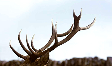
There is one type of mammal that engages in regenerative behavior in a very routine and reliable way, the deer. Male deer antlers regrow yearly as living tissue, with blood vessels and nerves wrapped around a fast growing boney structure. The researchers document a blastema-like structure present during antler regeneration, one similar to the structure involved in amphibian limb regeneration, suggesting a conserved biological feature available to vertebrate tissue regeneration.
There is another mammal with limited ability to regrow parts of a limb—mice. Mice can regenerate the tips of their foretoes. A cross-species comparison found that regenerative progenitor cells similar to those found in the deer antler blastema-like structure are also present in the mouse regenerative foretoe tip but not in nonregenerative mouse toes. These genes were also different from those found in axolotl limbs or zebrafish fins.
To fully document the gene transcription dynamics and asses cell type changes during antler regeneration, the research team pursued single-cell RNA sequencing of antlers at different stages of regeneration and a chromosome-level genome assembly of a male sika deer. 74,730 cells covering the critical stages of antler regeneration were analyzed, with some remarkable connections found between cell types reportedly crucial during limb regeneration in frogs and axolotl, as well as digit tip regeneration in mice.
An experiment was performed on mice to test the role of these progenitor cells. In the experiment, antler progenitor cells were introduced to the heads of laboratory mice. Antler-like boney cartilage formations appeared on the skull caps of mice that were not recruited from local tissues but entirely from the growth of transplanted stem cells, showing that the scientists had successfully isolated the essential cell types for regeneration.
News
Tumor “Stickiness” – Scientists Develop Potential New Way To Predict Cancer’s Spread
UC San Diego researchers have developed a device that predicts breast cancer aggressiveness by measuring tumor cell adhesion. Weakly adherent cells indicate a higher risk of metastasis, especially in early-stage DCIS. This innovation could [...]
Scientists Just Watched Atoms Move for the First Time Using AI
Scientists have developed a groundbreaking AI-driven technique that reveals the hidden movements of nanoparticles, essential in materials science, pharmaceuticals, and electronics. By integrating artificial intelligence with electron microscopy, researchers can now visualize atomic-level changes that were [...]
Scientists Sound Alarm: “Safe” Antibiotic Has Led to an Almost Untreatable Superbug
A recent study reveals that an antibiotic used for liver disease patients may increase their risk of contracting a dangerous superbug. An international team of researchers has discovered that rifaximin, a commonly prescribed antibiotic [...]
Scientists Discover Natural Compound That Stops Cancer Progression
A discovery led by OHSU was made possible by years of study conducted by University of Portland undergraduates. Scientists have discovered a natural compound that can halt a key process involved in the progression [...]
Scientists Just Discovered an RNA That Repairs DNA Damage – And It’s a Game-Changer
Our DNA is constantly under threat — from cell division errors to external factors like sunlight and smoking. Fortunately, cells have intricate repair mechanisms to counteract this damage. Scientists have uncovered a surprising role played by [...]
What Scientists Just Discovered About COVID-19’s Hidden Death Toll
COVID-19 didn’t just claim lives directly—it reshaped mortality patterns worldwide. A major international study found that life expectancy plummeted across most of the 24 analyzed countries, with additional deaths from cardiovascular disease, substance abuse, and mental [...]
Self-Propelled Nanoparticles Improve Immunotherapy for Non-Invasive Bladder Cancer
A study led by Pohang University of Science and Technology (POSTECH) and the Institute for Bioengineering of Catalonia (IBEC) in South Korea details the creation of urea-powered nanomotors that enhance immunotherapy for bladder cancer. The nanomotors [...]
Scientists Develop New System That Produces Drinking Water From Thin Air
UT Austin researchers have developed a biodegradable, biomass-based hydrogel that efficiently extracts drinkable water from the air, offering a scalable, sustainable solution for water access in off-grid communities, emergency relief, and agriculture. Discarded food [...]
AI Unveils Hidden Nanoparticles – A Breakthrough in Early Disease Detection
Deep Nanometry (DNM) is an innovative technique combining high-speed optical detection with AI-driven noise reduction, allowing researchers to find rare nanoparticles like extracellular vesicles (EVs). Since EVs play a role in disease detection, DNM [...]
Inhalable nanoparticles could help treat chronic lung disease
Nanoparticles designed to release antibiotics deep inside the lungs reduced inflammation and improved lung function in mice with symptoms of chronic obstructive pulmonary disease By Grace Wade Delivering medication to the lungs with inhalable nanoparticles [...]
New MRI Study Uncovers Hidden Lung Abnormalities in Children With Long COVID
Long COVID is more than just lingering symptoms—it may have a hidden biological basis that standard medical tests fail to detect. A groundbreaking study using advanced MRI technology has uncovered significant lung abnormalities in [...]
AI Struggles with Abstract Thought: Study Reveals GPT-4’s Limits
While GPT-4 performs well in structured reasoning tasks, a new study shows that its ability to adapt to variations is weak—suggesting AI still lacks true abstract understanding and flexibility in decision-making. Artificial Intelligence (AI), [...]
Turning Off Nerve Signals: Scientists Develop Promising New Pancreatic Cancer Treatment
Pancreatic cancer reprograms nerve cells to fuel its growth, but blocking these connections can shrink tumors and boost treatment effectiveness. Pancreatic cancer is closely linked to the nervous system, according to researchers from the [...]
New human antibody shows promise for Ebola virus treatment
New research led by scientists at La Jolla Institute for Immunology (LJI) reveals the workings of a human antibody called mAb 3A6, which may prove to be an important component for Ebola virus therapeutics. [...]
Early Alzheimer’s Detection Test – Years Before Symptoms Appear
A new biomarker test can detect early-stage tau protein clumping up to a decade before it appears on brain scans, improving early Alzheimer’s diagnosis. Unlike amyloid-beta, tau neurofibrillary tangles are directly linked to cognitive decline. Years [...]
New mpox variant can spread rapidly across borders
International researchers, including from DTU National Food Institute, warn that the ongoing mpox outbreak in the Democratic Republic of the Congo (DRC) has the potential to spread across borders more rapidly. The mpox virus [...]
