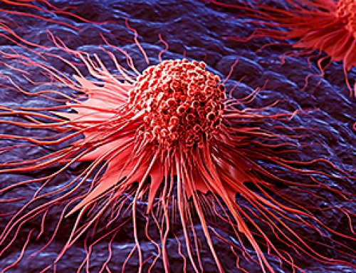
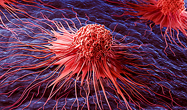
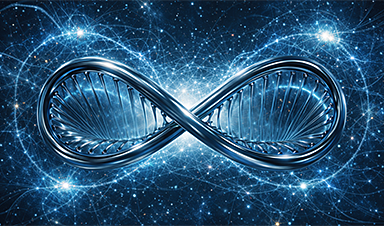
Scientists at EMBL have captured how human chromosomes fold into their signature rod shape during cell division, using a groundbreaking method called LoopTrace.
By observing overlapping DNA loops forming in high resolution, they revealed that large loops form first, followed by nested smaller loops, all repelling each other into compact structures. This new insight not only reshapes our understanding of chromosome mechanics but could also help explain errors that lead to cancer and genetic disorders.
The Mystery of Chromosome Division
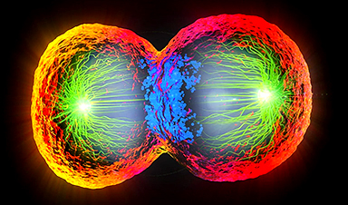
One of the most remarkable abilities of living cells is their capacity to divide, allowing organisms to grow, heal, and renew themselves. To do this, a cell must first make an exact copy of its DNA, its genome, and ensure each daughter cell receives a complete set.
In humans, that means carefully packaging 46 chromosomes and distributing them equally. Before division, each chromosome transforms into a compact, X-shaped structure made of two identical, rod-like copies. But exactly how cells manage to reshape and organize their DNA for this process has remained a mystery.
Now, for the first time, scientists at EMBL have directly visualized this process in high resolution using a new chromatin tracing technique. Their study reveals that during cell division, the long strands of DNA form a series of overlapping loops that push away from one another. This repulsion causes the loops to stack, ultimately giving each chromosome its characteristic rod-like shape.

Looping DNA to Shape Chromosomes
Scientists have long hypothesized the importance of DNA loops in building and maintaining chromosomal structure. First identified in the 1990s, condensins are large protein complexes that bind DNA during cell division and extrude it to create loops of varying sizes. Previous studies from EMBL have shed light on the structural mechanics of this process and their essential role in packing chromosomes into forms that can be easily moved between cells.
In fact, mutations in condensin structure can result in severe chromosome segregation defects and lead to cell death, cancer formation, or rare developmental disorders called 'condensinopathies'.
Solving the DNA Imaging Problem
"However, observing how this looping process occurs on the cellular scale and contributes to chromosome structure is challenging," said Andreas Brunner, postdoc in EMBL Heidelberg's Ellenberg Group and a lead author of the new paper. "This is because methods for visualizing DNA with high resolution are usually chemically harsh and require high temperatures, which together disrupt the native structure of DNA."
Kai Beckwith, a former postdoc in the Ellenberg Group and currently an associate professor at the Norwegian University of Science and Technology (NTNU), set out to solve this problem. Beckwith and colleagues used a method to gently remove one strand of DNA in cells at various stages of cell division, keeping the chromosome structure intact. They could then use targeted sets of DNA-binding labels to observe the nanoscale organization of this uncovered DNA strand. This technique, called LoopTrace, helped the researchers directly observe DNA in dividing cells as it progressively formed loops and folds.
"Andreas and I were now able to visualize the structure of chromosomes as they started to change shape," said Beckwith. "This was crucial for understanding how the DNA was folded by the condensin complexes."
Nested Loops and DNA Compaction
From their data, the scientists realized that during cell division, DNA forms loops in two stages. First, it forms stable large loops, which then subdivide into smaller, short-lived nested loops, increasing the compaction at each stage. Two types of condensin protein complexes enable this process.
To understand how this looping eventually gives rise to rod-shaped chromosomes, the researchers built a computational model based on two simple assumptions. First, as observed, DNA forms overlapping loops – first large and then small – across its length with the help of Condensins. Second, these loops repel each other due to their structure and the chemistry of DNA. When the scientists fed these two assumptions into their model, they found that this was sufficient to give rise to a rod-shaped chromosome structure.
Overlapping Loops Are Key
"We realized that these condensin-driven loops are much larger than previously thought, and that it was very important that the large loops overlap to a significant extent," said Beckwith. "Only these features allowed us to recapitulate the native structure of mitotic chromosomes in our model and understand how they can be segregated during cell division."
In the future, the researchers plan to study this process in more detail, especially to understand how additional factors, such as molecular regulators, affect this compaction process. In 2024, Jan Ellenberg and his team received funding of €3.1 million (~$3.4 million) as an ERC Advanced Grant, to study the folding principles of chromosomes during and following cell division.
A Milestone for Chromosome Biology
"Our newest paper published in the scientific journal Cell marks a milestone in our understanding of how the cell is able to pack chromosomes for their accurate segregation into daughter cells," said Jan Ellenberg, Senior Scientist at EMBL Heidelberg. "It will be the basis to understand the molecular mechanism of rescaling the genome for faithful inheritance and thus rationally predict how errors in this process that underlie human disease could be prevented in the future."
In the meantime, a second study from the Ellenberg Team, led by Andreas Brunner and recently published in the Journal of Cell Biology, shows that the nested loop mechanism is fundamental to the biology of cells, and continues during the cell's growth phase with another family of DNA loop forming protein complexes, called cohesins.
Looping Mechanisms Across Cell Phases
"We were surprised to find that the same core principle of sequential and hierarchical DNA loop formation is used to either tightly pack chromosomes during division into safely movable entities, or to unpack them afterward to read out the information they contain," said Ellenberg. "In the end, small, but key mechanistic differences, such as the non-overlapping nature of cohesin-driven loops compared to the strongly overlapping condensin-driven loops might be sufficient to explain the vast differences that we see in the shape the genome takes in interphase and mitosis under the microscope."
References:
Reference: "Nanoscale DNA tracing reveals the self-organization mechanism of mitotic chromosomes" by Kai Sandvold Beckwith, Andreas Brunner, Natalia Rosalia Morero, Ralf Jungmann and Jan Ellenberg, 24 March 2025, Cell.
DOI: 10.1016/j.cell.2025.02.028
"Quantitative imaging of loop extruders rebuilding interphase genome architecture after mitosis" by Andreas Brunner, Natalia Rosalía Morero, Wanlu Zhang, M. Julius Hossain, Marko Lampe, Hannah Pflaumer, Aliaksandr Halavatyi, Jan-Michael Peters, Kai S. Beckwith and Jan Ellenberg, 9 January 2025, Journal of Cell Biology.
DOI: 10.1083/jcb.202405169
News

Scientists Discover Stem Cells That Could Regrow Teeth and Bone
Scientists just uncovered the cellular “blueprint” that could one day let us regrow real teeth. Researchers at Science Tokyo have uncovered two distinct stem cell lineages that play a central role in forming tooth [...]
Scientists Uncover Fatal Weakness in “Zombie Cells” Linked to Cancer
A newly identified weakness in “zombie” cells may open the door to more precise cancer treatments by turning their own survival strategy against them. A new class of drugs takes advantage of a recently [...]

Bowel and Ovarian Cancers Are Dramatically Rising in Young Adults, Scientists Aren’t Sure Why
Cancer incidence is increasing, especially among younger adults, and current risk factors don’t fully account for the trend. Scientists suggest other underlying causes may be contributing. Cancer patterns in England are shifting in a [...]

New Immune Pathway Could Supercharge mRNA Cancer Vaccines
A surprising backup system in the immune response to mRNA vaccines may hold the key to more effective cancer treatments. The arrival of mRNA vaccines against SARS-CoV-2 in 2020 marked a turning point in the COVID-19 pandemic. Today, [...]
Scientists Discover “Molecular Switch” That Fuels Alzheimer’s Brain Inflammation
A newly identified trigger of brain inflammation could offer a fresh target for slowing Alzheimer’s progression. The brain has its own built-in immune system that identifies threats and responds to them. In Alzheimer’s disease, growing evidence [...]

Molecular Manufacturing: The Future of Nanomedicine – New book from NanoappsMedical Inc.
This book explores the revolutionary potential of atomically precise manufacturing technologies to transform global healthcare, as well as practically every other sector across society. This forward-thinking volume examines how envisaged Factory@Home systems might enable the cost-effective [...]
Forgotten Medicinal Plant Shows Promise in Fighting Dangerous Superbugs
A traditional medicinal plant, tormentil, shows promise against antibiotic-resistant bacteria in laboratory tests. Its compounds work by limiting bacterial growth and boosting antibiotic performance. Before the development of modern antibiotics, plant-based remedies were commonly [...]

NanoMedical Brain/Cloud Interface – Explorations and Implications. A new book from Frank Boehm
New book from Frank Boehm, NanoappsMedical Inc Founder: This book explores the future hypothetical possibility that the cerebral cortex of the human brain might be seamlessly, safely, and securely connected with the Cloud via [...]

New Research Finds Shocking Link Between Chili Peppers and Cancer
If you love spicy food, you are not alone. But scientists are taking a closer look at whether eating a lot of chili peppers could affect your cancer risk. Could your love of spicy [...]

New book from Nanoappsmedical Inc. – Global Health Care Equivalency
A new book by Frank Boehm, NanoappsMedical Inc. Founder. This groundbreaking volume explores the vision of a Global Health Care Equivalency (GHCE) system powered by artificial intelligence and quantum computing technologies, operating on secure [...]
Scientists Create “Neurobots” – Living Machines With Their Own Nervous Systems
Neurobots—xenobots with neurons—show self-organized nervous systems and enhanced behaviors, revealing new insights into how biology builds functional structures. In 2020, researchers at Tufts University developed tiny living structures known as xenobots using frog cells. These microscopic organisms [...]

Our books now available worldwide!
Online Sellers other than Amazon, Routledge, and IOPP Indigo Global Health Care Equivalency in the Age of Nanotechnology, Nanomedicine and Artifcial Intelligence Global Health Care Equivalency In The Age Of Nanotechnology, Nanomedicine And Artificial [...]

Amazonian Chocolate Could Become the Next Superfood, Scientists Say
New research into Amazonian cocoa reveals that its value may extend beyond flavor alone. Chocolate from the Amazon is already known worldwide for its distinctive taste, but new research suggests it may offer even [...]
Nanobody repairs misfolded CFTR inside cells, boosting function in cystic fibrosis
A tiny antibody component could fundamentally transform the treatment of cystic fibrosis: For the first time, researchers have succeeded in developing a so-called nanobody that penetrates directly into human cells and can repair the [...]

20-Year Study Finds Daily Multivitamins Don’t Extend Lifespan
A large, decades-long study of over 390,000 U.S. adults challenges a widespread assumption about daily multivitamins. Multivitamins are a daily habit for millions of Americans, often taken with the expectation that they will extend [...]
Novel Investment Paradigms for Regenerative Healthcare Ecosystems
Introduction The transition toward regenerative healthcare ecosystems—anchored in wellness optimization, disease prevention, eradication strategies, and healthy longevity—necessitates a structural reconfiguration of capital architectures, governance models, and incentive design. Regenerative healthcare, by definition, transcends episodic [...]