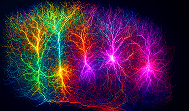
Artificial intelligence facilitates the visualization of neural connections in the brains of mice.
Scientists from Johns Hopkins have leveraged artificial intelligence to create a technique that allows for the visualization and monitoring of alterations in the strength of synapses — the connection points through which nerve cells in the brain communicate — in living organisms. The technique, as outlined in Nature Methods, could, according to the researchers, pave the way for an improved comprehension of how these connections in human brains evolve with learning, age, trauma, and disease.
“If you want to learn more about how an orchestra plays, you have to watch individual players over time, and this new method does that for synapses in the brains of living animals,” says Dwight Bergles, Ph.D., the Diana Sylvestre and Charles Homcy Professor in the Solomon H. Snyder Department of Neuroscience at the Johns Hopkins University (JHU) School of Medicine.
Bergles co-authored the study with colleagues Adam Charles, Ph.D., M.E., and Jeremias Sulam, Ph.D., both assistant professors in the biomedical engineering department, and Richard Huganir, Ph.D., Bloomberg Distinguished Professor at JHU and Director of the Solomon H. Snyder Department of Neuroscience. All four researchers are members of Johns Hopkins’ Kavli Neuroscience Discovery Institute.
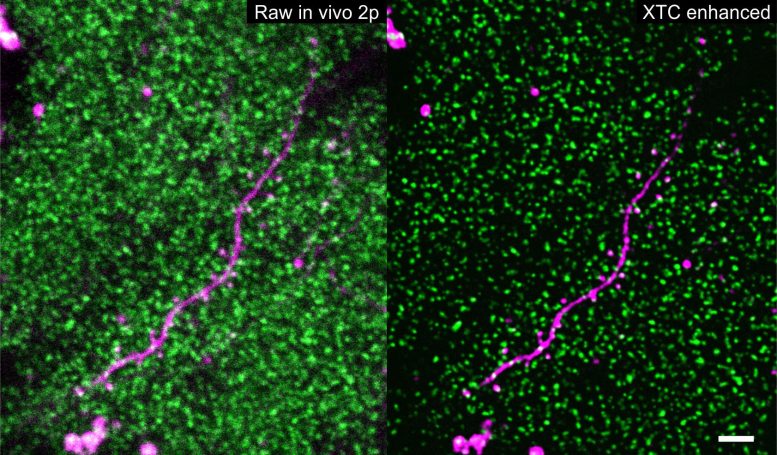
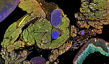
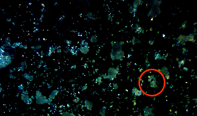
Thousands of SEP-GluA2 tagged synapses (green) surrounding a sparsely labeled dendrite (magenta) before and after XTC image resolution enhancement. Scale bar 5 microns. Credit: Xu, Y.K.T., Graves, A.R., Coste, G.I. et al. Nat Methods
Nerve cells transfer information from one cell to another by exchanging chemical messages at synapses (“junctions”). In the brain, the authors explain, different life experiences, such as exposure to new environments and learning skills, are thought to induce changes at synapses, strengthening or weakening these connections to allow learning and memory. Understanding how these minute changes occur across the trillions of synapses in our brains is a daunting challenge, but it is central to uncovering how the brain works when healthy and how it is altered by disease.
To determine which synapses change during a particular life event, scientists have long sought better ways to visualize the shifting chemistry of synaptic messaging, necessitated by the high density of synapses in the brain and their small size — traits that make them extremely hard to visualize even with new state-of-the-art microscopes.
“We needed to go from challenging, blurry, noisy imaging data to extract the signal portions we need to see,” Charles says.
To do so, Bergles, Sulam, Charles, Huganir, and their colleagues turned to machine learning, a computational framework that allows the flexible development of automatic data processing tools. Machine learning has been successfully applied to many domains across biomedical imaging, and in this case, the scientists leveraged the approach to enhance the quality of images composed of thousands of synapses. Although it can be a powerful tool for automated detection, greatly surpassing human speeds, the system must first be “trained,” teaching the algorithm what high-quality images of synapses should look like.
In these experiments, the researchers worked with genetically altered mice in which glutamate receptors — the chemical sensors at synapses — glowed green (fluoresced) when exposed to light. Because each receptor emits the same amount of light, the amount of fluorescence generated by a synapse in these mice is an indication of the number of synapses, and therefore its strength.
As expected, imaging in the intact brain produced low-quality pictures in which individual clusters of glutamate receptors at synapses were difficult to see clearly, let alone to be individually detected and tracked over time. To convert these into higher-quality images, the scientists trained a machine learning algorithm with images taken of brain slices (ex vivo) derived from the same type of genetically altered mice. Because these images weren’t from living animals, it was possible to produce much higher quality images using a different microscopy technique, as well as low-quality images — similar to those taken in live animals — of the same views.
This cross-modality data collection framework enabled the team to develop an enhancement algorithm that can produce higher-resolution images from low-quality ones, similar to the images collected from living mice. In this way, data collected from the intact brain can be significantly enhanced and able to detect and track individual synapses (in the thousands) during multiday experiments.
To follow changes in receptors over time in living mice, the researchers then used microscopy to take repeated images of the same synapses in mice over several weeks. After capturing baseline images, the team placed the animals in a chamber with new sights, smells, and tactile stimulation for a single five-minute period. They then imaged the same area of the brain every other day to see if and how the new stimuli had affected the number of glutamate receptors at synapses.
Although the focus of the work was on developing a set of methods to analyze synapse level changes in many different contexts, the researchers found that this simple change in environment caused a spectrum of alterations in fluorescence across synapses in the cerebral cortex, indicating connections where the strength increased and others where it decreased, with a bias toward strengthening in animals exposed to the novel environment.
The studies were enabled through close collaboration among scientists with distinct expertise, ranging from molecular biology to artificial intelligence, who don’t normally work closely together. But such collaboration, is encouraged at the cross-disciplinary Kavli Neuroscience Discovery Institute, Bergles says. The researchers are now using this machine learning approach to study synaptic changes in animal models of Alzheimer’s disease, and they believe the method could shed new light on synaptic changes that occur in other disease and injury contexts.
“We are really excited to see how and where the rest of the scientific community will take this,” Sulam says.
Reference: “Cross-modality supervised image restoration enables nanoscale tracking of synaptic plasticity in living mice” by Yu Kang T. Xu, Austin R. Graves, Gabrielle I. Coste, Richard L. Huganir, Dwight E. Bergles, Adam S. Charles and Jeremias Sulam, 11 May 2023, Nature Methods.
DOI: 10.1038/s41592-023-01871-6
News
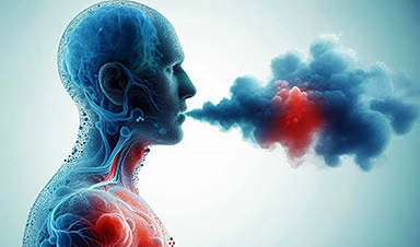
Studies detail high rates of long COVID among healthcare, dental workers
Researchers have estimated approximately 8% of Americas have ever experienced long COVID, or lasting symptoms, following an acute COVID-19 infection. Now two recent international studies suggest that the percentage is much higher among healthcare workers [...]
Melting Arctic Ice May Unleash Ancient Deadly Diseases, Scientists Warn
Melting Arctic ice increases human and animal interactions, raising the risk of infectious disease spread. Researchers urge early intervention and surveillance. Climate change is opening new pathways for the spread of infectious diseases such [...]
Scientists May Have Found a Secret Weapon To Stop Pancreatic Cancer Before It Starts
Researchers at Cold Spring Harbor Laboratory have found that blocking the FGFR2 and EGFR genes can stop early-stage pancreatic cancer from progressing, offering a promising path toward prevention. Pancreatic cancer is expected to become [...]
Breakthrough Drug Restores Vision: Researchers Successfully Reverse Retinal Damage
Blocking the PROX1 protein allowed KAIST researchers to regenerate damaged retinas and restore vision in mice. Vision is one of the most important human senses, yet more than 300 million people around the world are at [...]
Differentiating cancerous and healthy cells through motion analysis
Researchers from Tokyo Metropolitan University have found that the motion of unlabeled cells can be used to tell whether they are cancerous or healthy. They observed malignant fibrosarcoma cells and [...]
This Tiny Cellular Gate Could Be the Key to Curing Cancer – And Regrowing Hair
After more than five decades of mystery, scientists have finally unveiled the detailed structure and function of a long-theorized molecular machine in our mitochondria — the mitochondrial pyruvate carrier. This microscopic gatekeeper controls how [...]
Unlocking Vision’s Secrets: Researchers Reveal 3D Structure of Key Eye Protein
Researchers have uncovered the 3D structure of RBP3, a key protein in vision, revealing how it transports retinoids and fatty acids and how its dysfunction may lead to retinal diseases. Proteins play a critical [...]
5 Key Facts About Nanoplastics and How They Affect the Human Body
Nanoplastics are typically defined as plastic particles smaller than 1000 nanometers. These particles are increasingly being detected in human tissues: they can bypass biological barriers, accumulate in organs, and may influence health in ways [...]

Measles Is Back: Doctors Warn of Dangerous Surge Across the U.S.
Parents are encouraged to contact their pediatrician if their child has been exposed to measles or is showing symptoms. Pediatric infectious disease experts are emphasizing the critical importance of measles vaccination, as the highly [...]
AI at the Speed of Light: How Silicon Photonics Are Reinventing Hardware
A cutting-edge AI acceleration platform powered by light rather than electricity could revolutionize how AI is trained and deployed. Using photonic integrated circuits made from advanced III-V semiconductors, researchers have developed a system that vastly [...]
A Grain of Brain, 523 Million Synapses, Most Complicated Neuroscience Experiment Ever Attempted
A team of over 150 scientists has achieved what once seemed impossible: a complete wiring and activity map of a tiny section of a mammalian brain. This feat, part of the MICrONS Project, rivals [...]
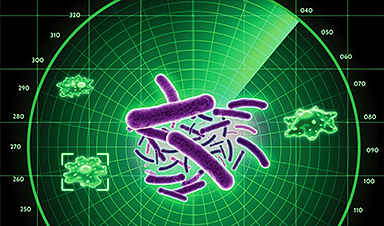
The Secret “Radar” Bacteria Use To Outsmart Their Enemies
A chemical radar allows bacteria to sense and eliminate predators. Investigating how microorganisms communicate deepens our understanding of the complex ecological interactions that shape our environment is an area of key focus for the [...]
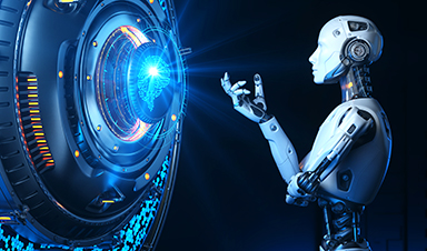
Psychologists explore ethical issues associated with human-AI relationships
It's becoming increasingly commonplace for people to develop intimate, long-term relationships with artificial intelligence (AI) technologies. At their extreme, people have "married" their AI companions in non-legally binding ceremonies, and at least two people [...]
When You Lose Weight, Where Does It Actually Go?
Most health professionals lack a clear understanding of how body fat is lost, often subscribing to misconceptions like fat converting to energy or muscle. The truth is, fat is actually broken down into carbon [...]
How Everyday Plastics Quietly Turn Into DNA-Damaging Nanoparticles
The same unique structure that makes plastic so versatile also makes it susceptible to breaking down into harmful micro- and nanoscale particles. The world is saturated with trillions of microscopic and nanoscopic plastic particles, some smaller [...]
AI Outperforms Physicians in Real-World Urgent Care Decisions, Study Finds
The study, conducted at the virtual urgent care clinic Cedars-Sinai Connect in LA, compared recommendations given in about 500 visits of adult patients with relatively common symptoms – respiratory, urinary, eye, vaginal and dental. [...]