Researchers have uncovered the 3D structure of RBP3, a key protein in vision, revealing how it transports retinoids and fatty acids and how its dysfunction may lead to retinal diseases.
Proteins play a critical role in the human body, acting as essential structural and functional components of cells, tissues, and organs. They are involved in a wide range of biological processes, from fundamental cellular functions such as DNA replication to more complex physiological activities, including those that enable vision.
Within the visual system, proteins are indispensable for detecting light, synthesizing photopigments in photoreceptor cells, and transmitting signals within these cells. Any disruption, whether through genetic mutation or protein malfunction, can impair normal vision and lead to a range of visual disorders.
Recently, scientists at the Institute of Physical Chemistry, Polish Academy of Sciences in collaboration with the International Centre for Translational Eye Research (ICTER) provided new structural insights into the RBP3 protein. Their findings have advanced our understanding of the visual cycle and its link to retinal diseases.
A Natural Optical Detector
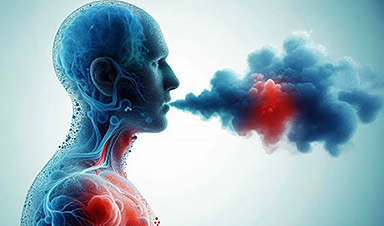
The human eye, our natural optical sensor, is a remarkably complex organ that enables us to perceive the world. Its function depends on the coordinated activity of numerous molecules. Vision begins in the retina, a thin layer of tissue lining the back of the eye, where light-sensitive cells known as photoreceptors (rods and cones) are located.
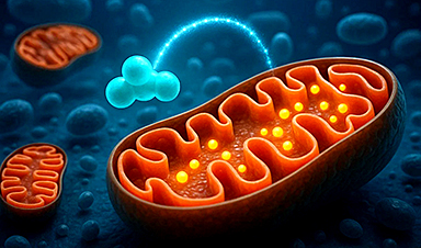
These photoreceptors detect light and convert it into electrical signals which are then transmitted to the brain via the optic nerve, allowing us to form visual images. A key molecule in this process is 11-cis-retinal (11cRAL), a light-sensitive compound that binds to opsin proteins such as rhodopsin. This interaction triggers the conversion of light into an electrical signal, initiating the visual process.
When photons are absorbed, a cascade of chemical reactions, including the isomerization of 11-cis-retinal (11cRAL) to all-trans-retinal, initiates vision. To enable continued vision, the 11cRAL must be continuously regenerated through a process called the visual cycle. Here the story begins…
Enter RBP3: The Retinoid Transporter
This is where another molecule enters the picture. That is Retinol-binding protein 3 (RBP3), a special protein located in the intercellular matrix that maintains the proper functioning of the visual cycle. RBP3 works as a transporter of retinoids between photoreceptors and retinal pigment epithelium cells and is also known to bind some important fatty acids. It shuttles crucial molecules back and forth from the photoreceptors making the visual pigments ready for the multiple reactions under the photons triggering.
The severity of diabetic retinopathy, an eye disease associated with diabetes, is associated with decreased levels of RBP3, and leads to progressive vision loss.
As RBP3 interacts with receptors like the glucose transporter 1 (GLUT1) and vascular endothelial growth factor (VEGF), been involved in blood vessel growth and cellular signaling in the eye. Disrupted RBP3 causes accumulation of retinal “waste products”, such as lipofuscin, which may cause oxidative damage to the RPE and photoreceptor cells. Besides diabetic retinopathy, RBP3 level disruption can also lead to retinitis pigmentosa, pan-retinal degeneration, and myopia.
Uncovering RBP3’s Structure
Although the RBP3 connection with these diseases is well known, the mechanisms of the binding to retinoids to transport them are still not satisfactorily described. This mystery intrigued the international team of researchers led by Dr. Humberto Fernandes from the Institute of Physical Chemistry, Polish Academy of Sciences – International Centre for Translational Eye Research (ICTER) to solve that mystery. They focused on the insight into the detailed structure of the RBP3 when it binds different retinoids and fatty acids.
The main aim of their investigations was to overcome the lack of an experimental structural model for the native form of RBP3. To achieve this, the authors purified the porcine RBP3 (pRBP3) and analyzed its structure using cryo-electron microscopy (cryoEM), where data was collected under cryogenic conditions, and after that data was refined by multiple steps and software to get the final 3D structure/model of the protein.
Additionally, small-angle X-ray scattering (SAXS) was used to provide data on the conformation changes depending on the cargo molecules. Interestingly, the structure of the RBP3 can be elongated, or bent, suggesting the dynamic changes in the structure while docking its cargo.
“Based on previous knowledge of RBP3 properties and straightforward methods for isolation of the porcine variant of RBP3, we purified porcine RBP3, and obtained a protein with Förster resonance energy transfer behaviour analogous to other RBP3s. Through analysis of cryoEM data, we determined a structure at 3.67 Å resolution of the porcine RBP3 protein and observed conformational changes upon ligand binding,” says Dr. Humberto Fernandes
Insights into RBP3 Function
Experimental results enabled the determination of the 3D structure and revealed conformational changes upon binding to its ligand as a step forward in the insight into the RBP3 functional mechanisms during the visual cycle.
RBP3 as a large molecule consisting of four retinoid-binding modules, has long lost its original catalytic functionality, and it evolved to be a cargo transporter interacting with a variety of molecules and delivering retinoids and fatty acids in the eye.
Research findings show the protein changes employing its shape during the binding of different molecules, which relates to the effectiveness of the interaction with the other molecules in the cargo and signaling. As a result, the conformational changes may play a significant role in the regulation of the light conversion into the visual signals.
Dr. Fernandes remarks, “In all measured parameters, the porcine variant mimics the more completely characterized bovine variant. The capacity of RBP3 to load different retinoids and fatty acids, the ability of the latter to displace the former, and the conformational changes dependent on ligand identity might be the basis for the loading and unloading of retinoids (and potentially DHA) to the intended cell types bordering the IPM intercellular matrix. Thus, RBP3 complexes merit further investigation.”
Understanding the proteins, including genetic mutations that affect the protein’s behaviour, like RBP3, is crucial to describe the mechanisms of the processes that appear in retinal diseases. Revealing the detailed structure of this bioactive molecule is a milestone in the studies on the interactions with different proteins.
The presented findings bring the bright light into potentially more effective and faster diagnostics, where the RBP3 molecule would work as an early-stage retinal disease development biomarker. What is more, it can help in the regulation of the RBP3 activity to develop treatments for the disruption of the visual process.
Reference: “CryoEM structure and small-angle X-ray scattering analyses of porcine retinol-binding protein 3” by Vineeta Kaushik, Luca Gessa, Nelam Kumar, Matyáš Pinkas, Mariusz Czarnocki-Cieciura, Krzysztof Palczewski, Jiří Nováček and Humberto Fernandes, 1 January 2025, Open Biology.
DOI: 10.1098/rsob.240180
News
This Tiny Cellular Gate Could Be the Key to Curing Cancer – And Regrowing Hair
After more than five decades of mystery, scientists have finally unveiled the detailed structure and function of a long-theorized molecular machine in our mitochondria — the mitochondrial pyruvate carrier. This microscopic gatekeeper controls how [...]
Unlocking Vision’s Secrets: Researchers Reveal 3D Structure of Key Eye Protein
Researchers have uncovered the 3D structure of RBP3, a key protein in vision, revealing how it transports retinoids and fatty acids and how its dysfunction may lead to retinal diseases. Proteins play a critical [...]

5 Key Facts About Nanoplastics and How They Affect the Human Body
Nanoplastics are typically defined as plastic particles smaller than 1000 nanometers. These particles are increasingly being detected in human tissues: they can bypass biological barriers, accumulate in organs, and may influence health in ways [...]

Measles Is Back: Doctors Warn of Dangerous Surge Across the U.S.
Parents are encouraged to contact their pediatrician if their child has been exposed to measles or is showing symptoms. Pediatric infectious disease experts are emphasizing the critical importance of measles vaccination, as the highly [...]
AI at the Speed of Light: How Silicon Photonics Are Reinventing Hardware
A cutting-edge AI acceleration platform powered by light rather than electricity could revolutionize how AI is trained and deployed. Using photonic integrated circuits made from advanced III-V semiconductors, researchers have developed a system that vastly [...]
A Grain of Brain, 523 Million Synapses, Most Complicated Neuroscience Experiment Ever Attempted
A team of over 150 scientists has achieved what once seemed impossible: a complete wiring and activity map of a tiny section of a mammalian brain. This feat, part of the MICrONS Project, rivals [...]
The Secret “Radar” Bacteria Use To Outsmart Their Enemies
A chemical radar allows bacteria to sense and eliminate predators. Investigating how microorganisms communicate deepens our understanding of the complex ecological interactions that shape our environment is an area of key focus for the [...]
Psychologists explore ethical issues associated with human-AI relationships
It's becoming increasingly commonplace for people to develop intimate, long-term relationships with artificial intelligence (AI) technologies. At their extreme, people have "married" their AI companions in non-legally binding ceremonies, and at least two people [...]
When You Lose Weight, Where Does It Actually Go?
Most health professionals lack a clear understanding of how body fat is lost, often subscribing to misconceptions like fat converting to energy or muscle. The truth is, fat is actually broken down into carbon [...]
How Everyday Plastics Quietly Turn Into DNA-Damaging Nanoparticles
The same unique structure that makes plastic so versatile also makes it susceptible to breaking down into harmful micro- and nanoscale particles. The world is saturated with trillions of microscopic and nanoscopic plastic particles, some smaller [...]
AI Outperforms Physicians in Real-World Urgent Care Decisions, Study Finds
The study, conducted at the virtual urgent care clinic Cedars-Sinai Connect in LA, compared recommendations given in about 500 visits of adult patients with relatively common symptoms – respiratory, urinary, eye, vaginal and dental. [...]
Challenging the Big Bang: A Multi-Singularity Origin for the Universe
In a study published in the journal Classical and Quantum Gravity, Dr. Richard Lieu, a physics professor at The University of Alabama in Huntsville (UAH), which is a part of The University of Alabama System, suggests that [...]

New drug restores vision by regenerating retinal nerves
Vision is one of the most crucial human senses, yet over 300 million people worldwide are at risk of vision loss due to various retinal diseases. While recent advancements in retinal disease treatments have [...]
Shingles vaccine cuts dementia risk by 20%, new study shows
A shingles shot may do more than prevent rash — it could help shield the aging brain from dementia, according to a landmark study using real-world data from the UK. A routine vaccine could [...]
AI Predicts Sudden Cardiac Arrest Days Before It Strikes
AI can now predict deadly heart arrhythmias up to two weeks in advance, potentially transforming cardiac care. Artificial intelligence could play a key role in preventing many cases of sudden cardiac death, according to [...]

NanoApps Medical is a Top 20 Feedspot Nanotech Blog
There is an ocean of Nanotechnology news published every day. Feedspot saves us a lot of time and we recommend it. We have been using it since 2018. Feedspot is a freemium online RSS [...]