Unprecedented views of the interior of cells and other nanoscale structures are now possible thanks to innovations in expansion microscopy. The advancements could help provide future insight into neuroscience, pathology, and many other biological and medical fields.
In the paper “Magnify is a universal molecular anchoring strategy for expansion microscopy,” published today (January 2, 2023) in the journal Nature Biotechnology, collaborators from Carnegie Mellon University, the University of Pittsburgh, and Brown University describe new protocols for dubbed Magnify.
“Magnify can be a potent and accessible tool for the biotechnology community,” said Yongxin (Leon) Zhao, the Eberly Family Career Development Associate Professor of Biological Sciences.
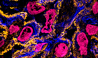
A video shows kidney cells. Expansion microscopy (ExM) provides unprecedented views of cell interiors. The emerging super-resolution imaging technique relies on physical — rather than optical — magnification. Advancements by Carnegie Mellon University’s Zhao Biophotonics Lab increases the expansion rate and allows many types of tissues to be viewed in 3D. Credit: Carnegie Mellon University
Magnify is a variant of expansion microscopy that allows researchers to use a new hydrogel formula, invented by Zhao’s team, that retains a spectrum of biomolecules, offers a broader application to a variety of tissues, and increases the expansion rate up to 11 times linearly or ~1,300 folds of the original volume.
“We overcame some of the longstanding challenges of expansion microscopy,” Zhao said. “One of the main selling points for Magnify is the universal strategy to keep the tissue’s biomolecules, including proteins, nucleus snippets, and carbohydrates, within the expanded sample.”
Zhao said that keeping different biological components intact matters because previous protocols required eliminating many various biomolecules that held tissues together. But these molecules could contain valuable information for researchers.
“In the past, to make cells really expandable, you need to use enzymes to digest proteins, so in the end, you had an empty gel with labels that indicate the location of the protein of interest,” he said. With the new method, the molecules are kept intact, and multiple types of biomolecules can be labeled in a single sample.
“Before, it was like having single-choice questions. If you want to label proteins, that would be the version one protocol. If you want to label nuclei, then that would be a different version,” Zhao said. “If you wanted to do simultaneous imaging, it was difficult. Now with Magnify, you can pick multiple items to label, such as proteins, lipids, and carbohydrates, and image them together.”
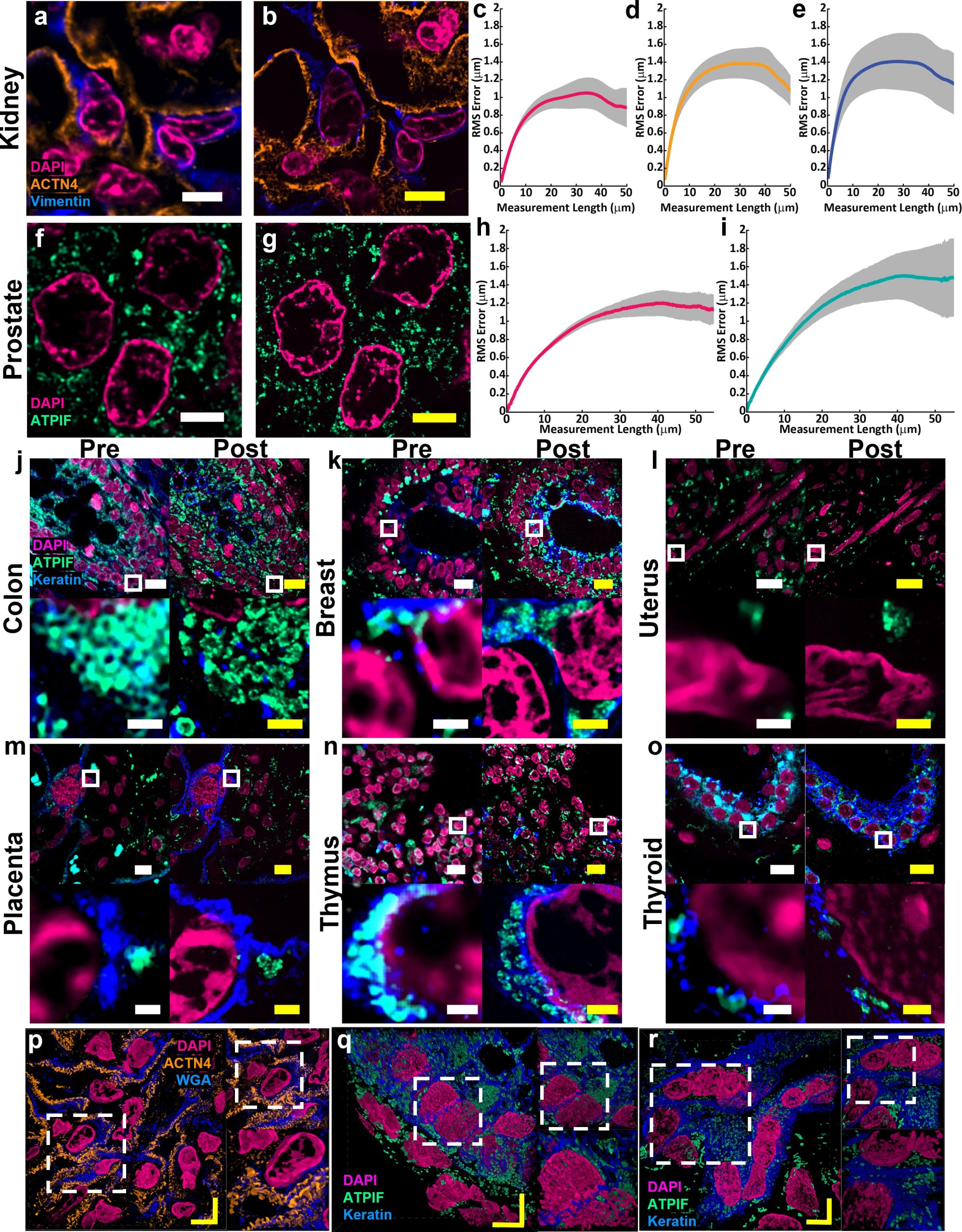
Example of (a) pre-expansion images of human kidney imaged at 60× and processed with SOFI compared to the same field of view (b) post-expansion with MAGNIFY taken at 40×. Magenta, DAPI; Orange, anti-alpha-actinin 4 (ACTN4); Blue, vimentin. Post expansion images are maximum intensity projected over 25 frames in z. (c-e) Root mean square (RMS) length measurement error as a function of measurement length for pre-expansion versus post expansion images for (c) DAPI, (d) ACTN4, and (e) Vimentin. Solid line, mean of channel; shaded area, standard error of mean (s.e.m); n = 5 technical replicates; average expansion factor, 8.64× (s.e.m 0.24). Example of (f) pre-expansion images of human prostate imaged at 60× and processed with SOFI compared to the same field of view (g) post-expansion with MAGNIFY taken at 40×. Magenta, DAPI; Green, Anti-ATPase Inhibitory Factor 1 (ATPIF). Post expansion images maximum intensity projected over 3 frames. (h-i) RMS length measurement error as a function of measurement length for pre-expansion versus post expansion images of (h) DAPI, and (i) ATPIF. Solid line, mean of channel; shaded area, s.e.m.; n = 4 technical replicates; average expansion factor, 10.38× (s.e.m 0.57). (j-o) Validation of MAGNIFY across multiple human tissue types. FFPE samples of human tissue were imaged at 40× (top left). Images were taken at 60×and processed with SOFI (bottom left). The white box indicates the field of view of the higher magnification images. The samples were then processed with the MAGNIFY protocol, and the same fields of view were imaged post-expansion in water at 10× (top right) and 40× (bottom right). Post expansion images were projected over 4-17 z slices. Magenta, DAPI; Green, ATPIF; Blue, Cytokeratin Pan Type I/II. Expansion factors in water were (j) Colon: 8.85×, (k) Breast: 9×, (l) Uterus: 8×, (m) Placenta: 8.75×, (n) Thymus: 10.00×, (o) Thyroid: 10.59×. (p-r) Example 3d images of human tissues: (p) kidney (Expansion factor 8.68×). Magenta, DAPI; Orange, ACTN4; Blue, WGA. (q) colon (Expansion factor 9.67×). Magenta, DAPI; Green, ATIPF; Blue, Cytokeratin Pan Type I/II. (r) Uterus (Expansion factor 8×). Magenta, DAPI; Green, ATIPF; Blue, Cytokeratin Pan Type I/II. Zoomed in regions indicated by dashed white box. Scale bars (yellow indicates post expansion images): (a) 5 μm; (b) 5 μm (physical scale post expansion: 40.75 μm; expansion factor: 8.15×); (f) 5 μm; (g) 5 μm (physical scale post expansion: 51.9 μm; expansion factor: 10.38×); (j-o) top: 10 μm; bottom: 1 μm; (p-t) 5 μm. Scale bars are all in biological scale. Credit: Courtesy of Carnegie Mellon University
Lab researchers Aleksandra Klimas, a postdoctoral researcher and Brendan Gallagher, a doctoral student, were first co-authors on the paper.
“This is an accessible way to image specimens in high resolution,” Klimas said. “Traditionally, you need expensive equipment and specific reagents and training. However, this method is broadly applicable to many types of sample preparations and can be viewed with standard microscopes that you would have in a biology laboratory.”
Gallagher, who has a background in neuroscience, said their goal was to make the protocols as compatible as possible for researchers who could benefit from adopting the Magnify as part of their toolkits.
“One of the key concepts that we tried to keep in mind was to meet researchers where they are and have them change as few things in their protocols as possible,” Gallagher said. “It works with different tissue types, fixation methods and even tissue that has been preserved and stored. It is very flexible, in that you don’t necessarily need to redesign experiments with Magnify in mind completely; it will work with what you have already.”
For researchers such as Simon Watkins, the founder and director of the Center for Biologic Imaging at the University of Pittsburgh and the Pittsburgh Cancer Institute, the fact that the new protocol is compatible with a broad range of tissue types — including preserved tissue sections — is important. For example, most expansion microscopy methods are optimized for brain tissue. In contrast, Magnify was tested on samples from various human organs and corresponding tumors including breast, brain and colon.
“Let’s say you have a tissue with dense and non-dense components, this gets around tissues that previously wouldn’t expand isometrically,” Watkins said. “Leon has been working hard on this to make this protocol work with tissues that have been archived.”
Xi (Charlie) Ren, an assistant professor of biomedical engineering at Carnegie Mellon, studies the lung tissue and how to model its morphogenesis and pathogenesis. Part of his research involves researching the motile cilia that function to clear mucus in the human conducting airway. At 200 nanometers in diameter and just a few micrometers in length, the structures are too small to see without time-intensive technology such as electron microscopy. Working in collaboration with Zhao’s lab, Ren’s team developed and delivered lung organoid models with specific defects in cilia ultrastructure and function to validate the ability of Magnify to visualize clinically relevant cilia pathology.
“With the latest Magnify techniques, we can expand those lung tissues and start to see some ultrastructure of the motile cilia even with a regular microscope, and this will expedite both basic and clinical investigations,” he said.
The researchers also were able to view defects in cilia in patient-specific lung cells known to have genetic mutations.
“The lung tissue engineering community always needs a better way to characterize the tissue system that we work with,” Ren said. He added that this work is an important first step and he hopes the collaborative work with Zhao’s lab will further be refined and applied to pathology samples found in tissue banks.
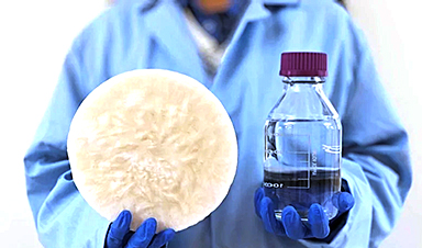
Finally, the hydrogel used in Magnify and developed in the Zhao lab is more robust than its predecessor, which was very fragile, causing breaks during the process.
“We are hoping to develop this technology to make it more accessible to the community,” he said. “There are different directions this can go. There’s a lot of interest in using this kind of tissue expansion technology for basic science.”
Alison Barth, the Maxwell H. and Gloria C. Connan Professor in the Life Sciences at Carnegie Mellon, studies synaptic connectivity during learning. She said the broad applications provided by the new methods will be a boon for researchers.
“The brain is a great place to take advantage of these super-resolution techniques,” said Barth, who collaborates with the Zhao Lab on several studies. “Microscopy methods will be beneficial for synaptic phenotyping and analysis across different brain conditions.
“One of the major advances in this paper is the method’s ability to work on many different types of tissue specimens.”
News
Shocking Amounts of Microplastics in the Brain – It Could Be Increasing Our Risk of Dementia
The brain has higher concentrations of plastic particles compared to other organs, with increased levels found in dementia patients. In a comprehensive commentary published in Brain Medicine, researchers highlight alarming new evidence of microplastic accumulation [...]

Baffling Scientists for Centuries: New Study Unravels Mystery of Static Electricity
ISTA physicists demonstrate that contact electrification depends on the contact history of materials. For centuries, static electricity has intrigued and perplexed scientists. Now, researchers from the Waitukaitis group at the Institute of Science and [...]
Tumor “Stickiness” – Scientists Develop Potential New Way To Predict Cancer’s Spread
UC San Diego researchers have developed a device that predicts breast cancer aggressiveness by measuring tumor cell adhesion. Weakly adherent cells indicate a higher risk of metastasis, especially in early-stage DCIS. This innovation could [...]

Scientists Just Watched Atoms Move for the First Time Using AI
Scientists have developed a groundbreaking AI-driven technique that reveals the hidden movements of nanoparticles, essential in materials science, pharmaceuticals, and electronics. By integrating artificial intelligence with electron microscopy, researchers can now visualize atomic-level changes that were [...]

Scientists Sound Alarm: “Safe” Antibiotic Has Led to an Almost Untreatable Superbug
A recent study reveals that an antibiotic used for liver disease patients may increase their risk of contracting a dangerous superbug. An international team of researchers has discovered that rifaximin, a commonly prescribed antibiotic [...]
Scientists Discover Natural Compound That Stops Cancer Progression
A discovery led by OHSU was made possible by years of study conducted by University of Portland undergraduates. Scientists have discovered a natural compound that can halt a key process involved in the progression [...]

Scientists Just Discovered an RNA That Repairs DNA Damage – And It’s a Game-Changer
Our DNA is constantly under threat — from cell division errors to external factors like sunlight and smoking. Fortunately, cells have intricate repair mechanisms to counteract this damage. Scientists have uncovered a surprising role played by [...]
What Scientists Just Discovered About COVID-19’s Hidden Death Toll
COVID-19 didn’t just claim lives directly—it reshaped mortality patterns worldwide. A major international study found that life expectancy plummeted across most of the 24 analyzed countries, with additional deaths from cardiovascular disease, substance abuse, and mental [...]
Self-Propelled Nanoparticles Improve Immunotherapy for Non-Invasive Bladder Cancer
A study led by Pohang University of Science and Technology (POSTECH) and the Institute for Bioengineering of Catalonia (IBEC) in South Korea details the creation of urea-powered nanomotors that enhance immunotherapy for bladder cancer. The nanomotors [...]
Scientists Develop New System That Produces Drinking Water From Thin Air
UT Austin researchers have developed a biodegradable, biomass-based hydrogel that efficiently extracts drinkable water from the air, offering a scalable, sustainable solution for water access in off-grid communities, emergency relief, and agriculture. Discarded food [...]

AI Unveils Hidden Nanoparticles – A Breakthrough in Early Disease Detection
Deep Nanometry (DNM) is an innovative technique combining high-speed optical detection with AI-driven noise reduction, allowing researchers to find rare nanoparticles like extracellular vesicles (EVs). Since EVs play a role in disease detection, DNM [...]
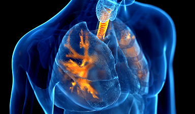
Inhalable nanoparticles could help treat chronic lung disease
Nanoparticles designed to release antibiotics deep inside the lungs reduced inflammation and improved lung function in mice with symptoms of chronic obstructive pulmonary disease By Grace Wade Delivering medication to the lungs with inhalable nanoparticles [...]
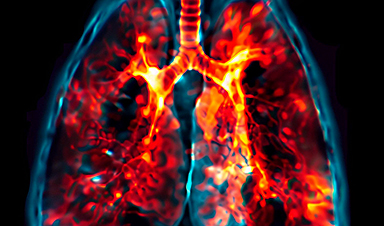
New MRI Study Uncovers Hidden Lung Abnormalities in Children With Long COVID
Long COVID is more than just lingering symptoms—it may have a hidden biological basis that standard medical tests fail to detect. A groundbreaking study using advanced MRI technology has uncovered significant lung abnormalities in [...]

AI Struggles with Abstract Thought: Study Reveals GPT-4’s Limits
While GPT-4 performs well in structured reasoning tasks, a new study shows that its ability to adapt to variations is weak—suggesting AI still lacks true abstract understanding and flexibility in decision-making. Artificial Intelligence (AI), [...]
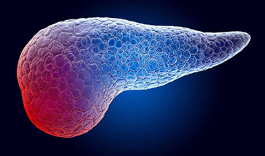
Turning Off Nerve Signals: Scientists Develop Promising New Pancreatic Cancer Treatment
Pancreatic cancer reprograms nerve cells to fuel its growth, but blocking these connections can shrink tumors and boost treatment effectiveness. Pancreatic cancer is closely linked to the nervous system, according to researchers from the [...]
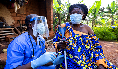
New human antibody shows promise for Ebola virus treatment
New research led by scientists at La Jolla Institute for Immunology (LJI) reveals the workings of a human antibody called mAb 3A6, which may prove to be an important component for Ebola virus therapeutics. [...]


